Topic: Alimentary, Hepatobiliary systems, Pancreas and SpleenSource: Internal
Explanation ready
Which of the following is not a derivative of the ventral mesentery?
Image not available for this question yet.
A) Falciform ligament
B) Coronary ligament
C) Lesser omentum
D) Gastrosplenic ligament
Correct Answer:D
Explanation:
The gastrosplenic ligament is a derivative of the dorsal mesentery.
Mesenteries are double layers of the peritoneum that enclose an organ and connect it to the body wall. They provide pathways for vessels, nerves, and lymphatics to and from the abdominal viscera.
The foregut gives rise to both the ventral and dorsal mesentery, whereas the midgut and hindgut give rise to only the dorsal mesentery. The ventral and dorsal mesenteries give rise to the following structures, as shown in the image below:
Q1352.
Anatomy
Medium
4m
Image missing
Topic: Alimentary, Hepatobiliary systems, Pancreas and SpleenSource: Internal
Explanation ready
Which of the following structures is not derived from the septum transversum?
Image not available for this question yet.
A) Falciform ligament
B) Ligamentum teres
C) Coronary ligament
D) Lesser omentum
Correct Answer:B
Explanation:
The ligamentum teres is not a derivative of the septum transversum, but a remnant of the left umbilical vein.
The septum transversum gives rise to:
Central tendon of the diaphragm
Connective tissue of the liver
Ventral mesentery of the foregut that has the following parts
Lesser omentum (ventral mesogastrium)
Ligaments of the liver (falciform ligament, triangular ligaments, and coronary ligaments)
Q1353.
Anatomy
Medium
4m
Image missing
Topic: Alimentary, Hepatobiliary systems, Pancreas and SpleenSource: Internal
Explanation ready
Which of the following structures does not develop in the mesogastrium?
Image not available for this question yet.
A) Liver
B) Spleen
C) Kidney
D) Pancreas
Correct Answer:C
Explanation:
The kidney is a retroperitoneal organ and does not develop in the mesogastrium. The mesenteries of the stomach include the ventral and dorsal mesogastrium.
Ventral mesogastrium (Lesser omentum), where the ventral pancreatic bud, liver, and gallbladder develop
Dorsal mesogastrium (Greater omentum), where the spleen, celiac trunk, and dorsal pancreatic bud develop
Q1354.
Anatomy
Medium
4m
Image missing
Topic: Alimentary, Hepatobiliary systems, Pancreas and SpleenSource: Internal
Explanation ready
The fibrous stroma of the liver is derived from which of the following structures?
Image not available for this question yet.
A) Foregut endoderm
B) Midgut endoderm
C) Hindgut endoderm
D) Septum transversum
Correct Answer:D
Explanation:
The fibrous stroma of the liver is derived from the septum transversum.
The liver develops as an outgrowth of the endodermal epithelium at the distal end of the foregut. This liver bud or hepatic diverticulum proliferates rapidly and penetrates the septum transversum.
The rapidly developing endodermal cells of the liver bud give rise to the parenchyma
(hepatocytes) and the lining of the biliary ducts. The endoderm-derived epithelial liver cells also intermingle with the vitelline and umbilical veins to form hepatic sinusoids.
The mesoderm of the septum transversum gives rise to the connective tissue, ligaments of the liver (except ligamentum teres), hematopoietic cells, and Kupffer cells.
The connection between the hepatic diverticulum and the foregut (duodenum) narrows, forming the bile duct.
The bile duct gives rise to a small ventral outgrowth called pars cystica that develops into the gallbladder and the cystic duct.
Q1355.
Anatomy
Medium
4m
Image missing
Topic: Alimentary, Hepatobiliary systems, Pancreas and SpleenSource: Internal
Explanation ready
Which of the following statements regarding the development of the pancreas is false?
Image not available for this question yet.
A) Inferior part of the head develops from the ventral pancreatic bud
B) Accessory duct develops from the dorsal pancreatic duct
C) Superior part of the head develops from the dorsal pancreatic bud
D) The uncinate process develops from the dorsal pancreatic bud
Correct Answer:D
Explanation:
The uncinate process develops from the ventral pancreatic bud.
The pancreas develops from two buds, dorsal and ventral, originating from the endodermal lining of the duodenum. When the duodenum rotates and becomes C-shaped, the ventral bud gets positioned immediately below and behind the dorsal bud. This is followed by the fusion of
the parenchyma and the duct systems of the dorsal and ventral pancreatic buds. The parenchyma of the pancreas:
Ventral pancreatic bud forms the lower part of the head and uncinate process.
Dorsal pancreatic bud forms the upper part of the head, neck, body, and tail.
Pancreatic ducts
The main pancreatic duct (of Wirsung) is formed by the duct of ventral bud (proximal part), oblique cross-communication between the 2 ducts (middle part), and the duct of dorsal bud (distal part).
The accessory pancreatic duct (of Santorini) is formed by the proximal part of the dorsal pancreatic duct.
The image given below shows the development of pancreas.
Q1356.
Anatomy
Medium
4m
Image missing
Topic: Alimentary, Hepatobiliary systems, Pancreas and SpleenSource: Internal
Explanation ready
Mesoderm of which of the following structures give rise to the spleen?
Image not available for this question yet.
A) Ventral mesogastrium
B) Dorsal mesogastrium
C) Dorsal mesoduodenum
D) Septum transversum
Correct Answer:B
Explanation:
The spleen develops from the mesoderm in the dorsal mesogastrium.
The mesenchymal cells in the dorsal mesogastrium form small lobular masses of splenic tissue called spleniculi. Later, these lobules fuse to form a single mass of the spleen. The capsule, septa, and connective tissue framework, including reticular fibers, develop from mesoderm. The mesenchymal cells also differentiate into lymphoblasts and other blood-forming cells.
When the spleen develops, the dorsal mesogastrium gets divided into an anterior segment called gastrosplenic ligament that extends from the spleen to stomach, and a posterior part called the lienorenal ligament that extends from the spleen to the left kidney.
Q1357.
Anatomy
Medium
4m
Image missing
Topic: Alimentary, Hepatobiliary systems, Pancreas and SpleenSource: Internal
Explanation ready
The rectum develops from
Image not available for this question yet.
A) Hindgut
B) Cloaca
C) Both a and
D) None of the above
Correct Answer:C
Explanation:
The rectum develops from both the hindgut and cloaca.
The section of the hindgut present caudal to the attachment of the allantoic diverticulum is called the cloaca.
The proximal part of the rectum develops from the hindgut
The distal part of the rectum develops from the dorsal part of the cloaca
Q1358.
Anatomy
Medium
4m
Image missing
Topic: Face, Nose Palate, Eye, EarSource: Internal
Explanation ready
Which of the following is not an embryologic structure that contributes to the formation of the face?
Image not available for this question yet.
A) Frontonasal prominence
B) Zygomatic prominence
C) Maxillary prominence
D) Mandibular prominence
Correct Answer:B
Explanation:
Zygomatic prominence is not an embryological structure that contributes to the formation of the face.
The face is derived from the following structures around the stomatodeum (primitive mouth):
Mandibular prominences and maxillary prominences of the first arch
Frontonasal prominence formed by the proliferation of mesenchyme ventral to the brain vesicles. It also gives rise to medial and lateral nasal prominences.
Structures contributing to th e development of face
Prominence Frontonasal
Medial nasal
Lateral nasal Maxillary
Mandibular
Structure formed
Forehead, bridge of the nose, also forms the medial and la teral nasal prominences
Philtrum of the upper lip, the crest of the nose, and the tip of the nose
Alae of nose
Upper part of cheeks, the late ral portion of the upper lip
Lower part of cheeks, chin, lo wer lip
Q1359.
Anatomy
Medium
4m
Image missing
Topic: Face, Nose Palate, Eye, EarSource: Internal
Explanation ready
What is the upper lip derived from?
Image not available for this question yet.
A) Maxillary prominence and mandibular prominence
B) Maxillary prominence and medial nasal prominence
C) Maxillary prominence and lateral nasal prominence
D) Mandibular prominence and medial nasal prominence
Correct Answer:B
Explanation:
The upper lip is formed by the fusion of the two maxillary prominences with the two medial nasal prominences.
Initially, two thickenings called nasal placodes are formed in the frontonasal prominence. The nasal placodes get depressed to become nasal pits. Each nasal pit has a ridge of tissue on the outer edge called lateral nasal prominence and on the inner edge called medial nasal prominence.
The 2 maxillary prominences on the lateral part of the stomodeum increase in size and grow medially. They compress the medial nasal prominences toward the midline. Later, the 2 maxillary prominences fuse with the 2 medial nasal prominences to form the upper lip.
Q1360.
Anatomy
Medium
4m
Image missing
Topic: Face, Nose Palate, Eye, EarSource: Internal
Explanation ready
From which structures do the alae of the nose develop?
Image not available for this question yet.
A) Frontonasal process
B) Medial nasal process
C) Lateral nasal process
D) Bucconasal membrane
Correct Answer:C
Explanation:
The alae of the nose develop from the lateral nasal processes.
The development of various components of the nose can be summarized as follows:
Bridge - The frontonasal process
Dorsum and tip - Fused medial nasal processes
Alae - The lateral nasal processes
Anterior nares (nostrils) - The original site of the nasal pit
Nasal cavity - The nasal sacs formed by the elongation of nasal pits
Topic: Face, Nose Palate, Eye, EarSource: Internal
Explanation ready
An infant underwent palatoplasty for bilateral complete cleft palate. This condition occurs due to:
Image not available for this question yet.
A) Non-fusion of palatine processes with premaxilla only
B) Non-fusion of palatine processes in midline only
C) Non-fusion of palatine process with premaxilla and with each other
D) Non-fusion of median nasal process with maxillary process
Correct Answer:C
Explanation:
Bilateral complete cleft palate is due to the non-fusion of palatine processes with the premaxilla and with each other.
The palate is formed from two main structures:
Primary palate: The small triangular anterior part of the palate is formed by the fused medial nasal processes (intermaxillary segment or premaxilla).
Secondary palate: The main part of the palate is formed by the 2 palatine processes that converge from the maxillary processes.
The cleft palate can occur in the primary palate (anterior cleft) or in the secondary palate
(posterior palate).
Complete cleft palate involves both the primary and secondary palate.
Incomplete cleft palate involves only the secondary palate.
In bilateral complete cleft palate, a Y-shaped cleft is seen with gaps between the primary and secondary palate and between the two halves of the secondary palate. In addition, there is a bilateral cleft upper lip.
Q1362.
Anatomy
Medium
4m
Image missing
Topic: Face, Nose Palate, Eye, EarSource: Internal
Explanation ready
A 1-week-old infant was diagnosed with congenital nasolacrimal duct obstruction. This structure is derived from which layer?
Image not available for this question yet.
A) Ectoderm
B) Mesoderm
C) Endoderm and ectoderm
D) Ectoderm and mesoderm
Correct Answer:A
Explanation:
The nasolacrimal duct is derived from the ectoderm of the nasolacrimal groove (also known as naso-optic furrow).
The deep furrow between the maxillary and lateral nasal prominence is known as the nasolacrimal groove.
The ectoderm in the floor of this groove becomes a solid epithelial cord and detaches from the overlying ectoderm.
This solid cord then canalizes to form the nasolacrimal duct.
The lower end of the duct acquires a connection to the nasal cavity.
The upper end of the duct widens to form the lacrimal sac.
Q1363.
Anatomy
Medium
4m
Image ready
Topic: Face, Nose Palate, Eye, EarSource: Internal
Explanation ready
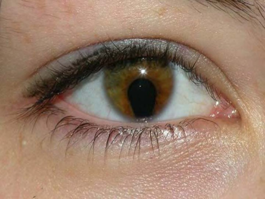
In an ophthalmology camp, you notice the following finding in a woman who had come for a routine checkup. It was diagnosed as coloboma of the iris. Which of the following mechanisms during embryonic development will most likely lead to the given condition? 134
A) Congenital detachment of the retina
B) Cavitation of the posterior chamber
C) Abnormal neural crest formation
D) Failure of closure of the choroid fissure
Correct Answer:D
Explanation:
The condition coloboma of the iris occurs due to failure of the choroid fissure to close in the distal part forming a cleft.
There is a small gap in the inferior surface of the optic cup known as the choroid fissure. This fissure allows the hyaloid vessels to reach the inner chamber of the eye. During the normal development of the eye, the distal part of the hyaloid artery degenerates. The choroid fissure then fuses and this makes the mouth of the optic cup into a round opening (the future pupil).
Failure of fusion of choroid fissure causes clefts in the eye known as coloboma.
If the failure to fuse is in the distal part, it can result in coloboma of the iris.
If the failure of fusion occurs in the proximal part, it can lead to coloboma of the retina/
choroid/
optic nerve, depending on the location of the defect.
Q1364.
Anatomy
Medium
4m
Image missing
Topic: Face, Nose Palate, Eye, EarSource: Internal
Explanation ready
Which of the following structures of the ear develops from all the three germ layers?
Image not available for this question yet.
A) Ear ossicles
B) External acoustic meatus
C) Middle ear
D) Tympanic membrane
Correct Answer:D
Explanation:
The tympanic membrane is derived from all three germ layers.
The tympanic membrane is formed by the apposition of the tubotympanic recess of the 1st pharyngeal pouch with the 1st pharyngeal cleft.
The outer layer of the tympanic membrane is derived from the ectoderm at the bottom of the external auditory meatus (1st pharyngeal cleft).
The inner layer of the tympanic membrane is derived from the endoderm lining the tympanic cavity (1st pharyngeal pouch).
The middle layer of the tympanic membrane is connective tissue derived from the mesoderm.
Q1365.
Anatomy
Medium
4m
Image missing
Topic: Face, Nose Palate, Eye, EarSource: Internal
Explanation ready
135 The tubotympanic recess gives rise to which structure?
Image not available for this question yet.
A) Auditory tube
B) External auditory meatus
C) Internal auditory meatus
D) Facial canal
Correct Answer:A
Explanation:
The tubotympanic recess forms the auditory tube. Development of the Middle Ear:
The distal part of the 1st pharyngeal pouch becomes the tubotympanic recess. The tubotympanic recess gives rise to the primitive tympanic cavity distally, while the proximal part remains narrow and forms the auditory tube (eustachian tube). It also forms the lining of the middle ear cavity.
The 3 ossicles are formed from mesenchymal condensations from the 1st pharyngeal arch
(malleus and incus) and 2nd pharyngeal arch (stapes).
• The tympanic membrane is formed where the ectoderm of the 1st pharyngeal cleft dips to meet the endoderm of the 1st pharyngeal pouch, with contribution from the intervening mesoderm.
Q1366.
Anatomy
Medium
4m
Image missing
Topic: Face, Nose Palate, Eye, EarSource: Internal
Explanation ready
The membranous labyrinth develops from which structure?
Image not available for this question yet.
A) First pharyngeal pouch
B) First pharyngeal cleft
C) Otic vesicle
D) Meckel's cartilage
Correct Answer:C
Explanation:
The membranous labyrinth of the internal ear is derived from the otic vesicle.
The surface ectoderm on either side of the hindbrain thickens to form otic placodes. The otic placodes invaginate to form otic vesicles (auditory vesicles).
The otic vesicle further divides into 2 components that develop into structures of the membranous labyrinth:
The ventral component forms the saccule and cochlear duct
The dorsal component forms the utricle, semicircular canals, and endolymphatic duct
Q1367.
Anatomy
Medium
4m
Image missing
Topic: Nervous System and Endocrine GlandsSource: Internal
Explanation ready
What is the spinal cord developed from?
Image not available for this question yet.
A) Neural tube
B) Notochord
C) Primitive streak
D) Rhombencephalon
Correct Answer:A
Explanation:
The spinal cord develops from the caudal part of the neural tube.
The nervous system develops from the neurectoderm i.e. the specialized ectoderm overlying the notochord. The neuroectoderm initially thickens to form the neural plate. The neural plate is then converted to a neural groove, followed by a neural tube.
The enlarged cranial part of neural tube forms the brain. It has 3 primary vesicles that form:
Forebrain (prosencephalon)
Midbrain (mesencephalon)
Hindbrain (rhombencephalon)
The narrow caudal part of the neural tube forms the spinal cord.
Q1368.
Anatomy
Medium
4m
Image missing
Topic: Nervous System and Endocrine GlandsSource: Internal
Explanation ready
The neuropore facilitates communication between the neural tube and
Image not available for this question yet.
A) Amniotic cavity
B) Yolk sac
C) Allantoic cavity
D) Uterus
Correct Answer:A
Explanation:
The neuropores facilitate communication between the neural tube and the amniotic cavity.
The neural plate first becomes tubular in the middle. The cranial and caudal ends of the neural tube remain open and are called the anterior and posterior neuropores, respectively.
The neuropores facilitate communication between the neural tube and the amniotic
cavity. Amniotic fluid provides nutrition to the developing neuroectodermal cells before the circulatory system is established.
Q1369.
Anatomy
Medium
4m
Image missing
Topic: Nervous System and Endocrine GlandsSource: Internal
Explanation ready
What is the adult representation of the location of anterior neuropore?
Image not available for this question yet.
A) Septum transversum
B) Terminal ventricle
C) Lamina terminalis
D) Foramen of Munro
Correct Answer:C
Explanation:
The location of the anterior neuropore is represented in adults as lamina terminalis.
The anterior neuropore closes around the 25th day of intrauterine life. Its location in adults is represented by the lamina terminalis. The lamina terminalis forms the anterior boundary of the third ventricle.
The posterior neuropore closes around the 28th day of intrauterine life. Its location in adults is represented by the terminal ventricle. The terminal ventricle is present in the proximal 5 to 6 mm of filum terminale.
Q1370.
Anatomy
Medium
4m
Image missing
Topic: Nervous System and Endocrine GlandsSource: Internal
Explanation ready
Which of the following statements best describes the sulcus limitans?
Image not available for this question yet.
A) It is found in the interpeduncular fossa
B) It is located between the alar and basal plates
C) It separates the medulla from the pons
D) It separates the hypothalamus from the thalamus
Correct Answer:B
Explanation:
The sulcus limitans is located between the sensory alar and the motor basal plates.
The cells in the wall of the neural tube proliferate and are subdivided into the inner mantle layer and outer marginal layer. Due to the fast growth of the mantle layer cells, the lumen of the neural tube gets compressed.
The line separating the compressed ventral part from the dorsal part is called the sulcus limitans. When the sulcus limitans is formed, the tube is divided into the dorsal or alar lamina and ventral or basal lamina.
In the spinal cord, the ventral thickenings or basal plates contain ventral motor horn cells. The dorsal thickenings i.e. the alar plates contain the interneurones that receive the terminals of primary sensory neurons.
Which of the following brainstem nuclei is not derived from the alar plate?
Image not available for this question yet.
A) Inferior olivary nucleus
B) Hypoglossal nucleus
C) Dentate nucleus
D) Mesencephalic nucleus
Correct Answer:B
Explanation:
The hypoglossal nucleus is not derived from the alar plate. The hypoglossal nucleus is a motor nucleus derived from the basal plate.
Embryologically, the continuous addition of neuroblasts to the mantle layer of the neural tube produces thickenings ventrally and dorsally - producing the basal and alar plates respectively. A longitudinal groove, called sulcus limitans, marks the boundary between the two.
Basal Plate Ventralthickening
Give rise tomotor nuclei-gene ral visceral efferent nuclei, sp ecial visceral efferent of CN I X, X, and XII; and somatic ef ferent of XII
Forms the motor part of the s pinal cord (ventral horns)
Alar plate Dorsalthickenings
Give rise tosensory nucleiCN V, VII, VIII, IX, X, inferior ol ivary nucleus, and mesencep halic nucleus of CN V
Forms the sensory part of the spinal cord (dorsal horns).T he rhombic lip develops into the cerebellum and its nuclei.
Q1372.
Anatomy
Medium
4m
Image missing
Topic: Nervous System and Endocrine GlandsSource: Internal
Explanation ready
Which of the following is the first commissure to develop?
Image not available for this question yet.
A) Fornix
B) Anterior commissure
C) Corpus callosum
D) Posterior commissure
Correct Answer:B
Explanation:
The first commissure to develop is the anterior commissure.
Commissures are fibre bundles that cross the midline and connect the right and left brain hemispheres. Many important fiber bundles cross at lamina terminalis.
Commissures at lamina terminalis:
Anterior commissure - first commissure to appear. These fibers connect the olfactory bulb and related brain areas to the corresponding structures in the contralateral hemisphere.
Hippocampal commissure (fornix commissure) - develops second. The fibers emerge from the hippocampus and reach the lamina terminalis. From here, they arch outside the choroid fissure to the mamillary body and the hypothalamus.
Corpus callosum - appears by the 10th week. It connects the non-olfactory areas of the right and the left cerebral cortices.
Commissures at other areas are:
Posterior commissure
Habenular commissure
Optic chiasma
The image given below shows the anterior commissure:
Q1373.
Anatomy
Medium
4m
Image ready
Topic: Nervous System and Endocrine GlandsSource: Internal
Explanation ready
Where does the structure marked as A develop from?
A) Basal lamina
B) Alar lamina
C) Lamina terminalis
D) Basal plate
Correct Answer:C
Explanation:
The structure marked as A in the image is the corpus callosum. It develops from the lamina terminalis.
Lamina terminalis is a part of the wall of the neural tube that closes the cranial end of the prosencephalon. The lamina terminalis becomes thickened to form the commissural plate that facilitates the passage of neurons between the two hemispheres.
There are 3 commissures that pass through lamina terminalis:
Anterior commissure
Hippocampal commissure
Corpus callosum
The corpus callosum is the largest of these commisures. Initially, corpus callosum forms a small bundle in the lamina terminalis. Due to the continuous expansion, it extends anteriorly and posteriorly arching over the thin roof of the diencephalon. It connects the non-olfactory areas of the right and the left cerebral cortices.
Q1374.
Anatomy
Medium
4m
Image missing
Topic: Nervous System and Endocrine GlandsSource: Internal
Explanation ready
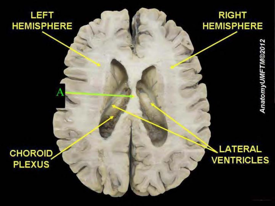
The septum pellucidum extends between:
Image not available for this question yet.
A) Anterior commissure and posterior commissure
B) Anterior commissure and corpus callosum
C) Anterior commissure and the fornix
D) Fornix and corpus callosum
Correct Answer:D
Explanation:
Septum pellucidum extends between the corpus callosum and the fornix.
The septum pellucidum is a membrane separating the anterior horns of the left and right lateral ventricles of the brain. It extends from the corpus callosum to the fornix.
It develops from the lamina terminalis. The part of the lamina terminalis that stretches between corpus callosum and fornix persists as septum pellucidum.
The image given below shows the septum pellucidum.
Q1375.
Anatomy
Medium
4m
Image missing
Topic: Nervous System and Endocrine GlandsSource: Internal
Explanation ready
Which of the following neural tube defects is characterized by gaps in the vertebral arches?
Image not available for this question yet.
A) Encephalocele
B) Spina bifida
C) Hydrancephaly
D) Syringomyelia
Correct Answer:B
Explanation:
Spina bifida is characterized by gaps in the vertebral arches.
Spina bifida is a general term used for neural tube defects affecting the spinal region and associated with splitting of the vertebral arches. It may or may not involve the underlying neural tissue.
There are 3 types of spina bifida, as shown in the image below:
Rachischisis is the most severe type of neural tube defect, where the entire neural tube fails to close. Rachischisis can occur in the spinal cord or brain regions of the neural tube. The exposed neural tissues often become necrotic.