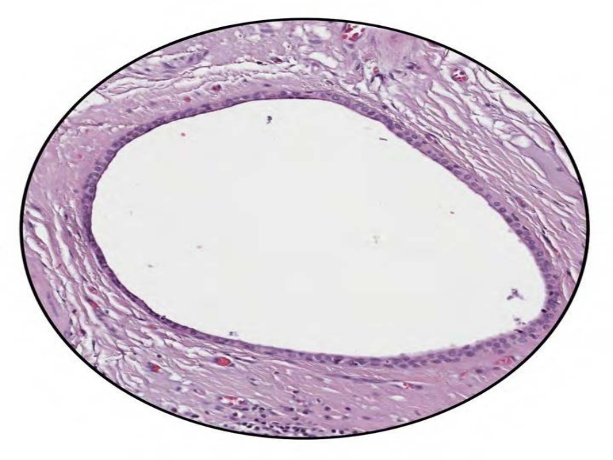
Given below is the histological section showing the excretory duct of the salivary gland. Which of the following ducts of the glands do not have the same lining epithelium? 182
A) Sweat gland
B) Sebaceous gland
C) Salivary gland
D) Exocrine pancreas
Correct Answer:B
Explanation:
The ducts of the sebaceous glands are lined by keratinised stratified squamous epithelium, whereas the ducts of sweat glands, salivary glands, and exocrine pancreas are lined by stratified cuboidal epithelium.
Microvilli are absent in collecting ducts. They are lined by simple columnar epithelium without microvilli.
The simple cuboidal or columnar epithelium may undergo certain specializations such as:
Microvilli - long finger-like projections arising from the surface of the epithelium, which increases the surface area of absorption. If the microvilli are long and are arranged regularly, parallel to one another, it gives a striated appearance on light microscopy. However, if the microvilli are arranged irregularly, it gives a brush border appearance. Microvilli are located at:
Small intestine - columnar cells with striated microvilli.
Gall bladder - columnar cells with brush border microvilli.
Proximal convoluted tubule - cuboidal cells with brush border microvilli
Stereocilia - long and branching microvilli. Unlike the cilia, the stereocilia are non-motile and hence, the preferred name is stereovilli. They are located at:
Epididymis (increase the surface area of absorption)
Cochlear and vestibular receptor cells (function as sensory transducers)
Cilia - hair-like projections on the epithelial cells that are concerned with the movement. The ciliated columnar epithelium is seen in the lining of the following structures:
Respiratory tract except in lower pharynx and vocal folds.
Tympanic cavity and auditory tube.
Uterine tube and efferent ductules of the testis.
Secretory - columnar cells may be specialized for secretions where they are referred to as glandular cells. Such cells form the mucin secreting and chief cells of the gastric epithelium.
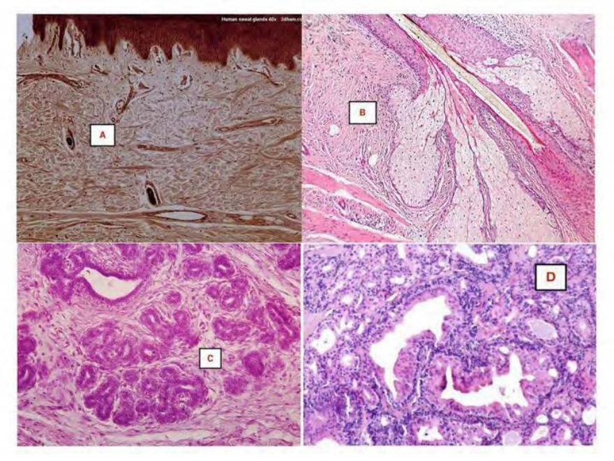
The exocrine glands are classified into 3 types, depending on the mode of secretion:
Merocrine glands - They are also known as eccrine or epicrine glands, in which the secretions are released out of the cell through a process called exocytosis with the cell remaining intact. The examples of such glands are:
Eccrine sweat glands
Salivary glands
Exocrine pancreas
Secretion of milk protein casein by mammary glands
Holocrine glands - In these glands, the entire cell disintegrates while discharging its
secretion. It is typically seen in sebaceous glands.
Apocrine glands - In these glands, the apical parts of the cells are shed off to discharge the secretion. Examples of apocrine glands are
Which of the following fibers of the connective tissue are stained black with silver impregnation? 185
Image not available for this question yet.
A) Type I collagen fibers
B) Elastic fibers
C) Reticular fibers
D) Type II collagen fibers
Correct Answer:C
Explanation:
Reticular fibers are stained black with silver impregnation method. Reticular fibers are also called argentophil fibers, due to their specific affinity for silver salts.
The fibers of connective tissue are of three main types. These include:
Collagen fibers
Reticular fibers
Elastic fibers
The staining characters of each of these fibers are as follows:
The image below shows reticular fibers in reticular connective tissue, stained black with silver impregnation method.
Connective tissue fibers
Collagen fibers
Verhoeff-Van Gieson
Hematoxylin and eosin
Silver impregnation
Masson's trichrome
Reticular fibers
Elastic fibers Aldehyde fuchsin
Stain
Unaided dye Light pink Blue
Red Brown
Silver impregnatio n
Verhoeff-Van Gies on
Purple
Appeara nce
White
Black Black
Connective tissue fibers Stain Appeara nce
Orcein Purplish brown
The images below show specimens of glans penis showing collagen fibers (Cf) stained blue and muscle fibers (Mf) stained red using Masson trichrome stain, and elastic fibers (Ef) stained black using Verhoeff-Van Gieson stain.
Sudan III and Sudan IV dyes are used for the staining of .
Image not available for this question yet.
A) Elastic tissue
B) Mucoid tissue
C) Adipose tissue
D) Reticular tissue
Correct Answer:C
Explanation:
Sudan III and Sudan IV dyes are used for the staining of adipose tissue. The image below shows adipose tissue stained with Sudan dye.
However, during the routine preparation of slides, the tissues are treated with fat solvents (like xylene or benzene), which dissolve the fat. Therefore, in such preparations, fat cells look like rounded empty spaces, as shown in the image below.
Which of the following statements is true regarding the constituents of bone matrix?
Image not available for this question yet.
A) The inorganic calcium salts make the bone tough and resilient.
B) The organic connective tissue affords resistance against compressive and impact forces.
C) Calcium phosphate is the major constituent of the inorganic salts present in the bone.
D) The organic matrix constitutes about 65 of the dry weight of the bone.
Correct Answer:C
Explanation:
Calcium phosphate in the form of calcium hydroxyapatite is the major constituent of the inorganic salts present in the bone.
The ground substance of the bone (bone matrix) is predominantly made up of inorganic mineral salts, which constitute about 60-70 of the dry weight of the bone. These inorganic salts are deposited within the organic matrix.
Inorganic mineral salts - makes the bone hard and rigid, and thus provides resistance to the compressive and impact forces as in weight-bearing or jumping respectively. Its constituents are:
Calcium phosphate in the form of calcium hydroxyapatite (85 of total salts)
Calcium carbonate (10 of total salts), magnesium, chloride, fluoride, citrate, sodium, and potassium.
Organic matrix - makes the bone tough and resilient (flexible) and thus provides resistance to the tensile forces. Its constituents are:
Type I collagen (predominantly)
Glycosaminoglycans, proteoglycans, and water.
Osteonectin and osteocalcin - predominant glycoproteins present in bone. These bind to calcium and help in the mineralization of the bone.
Other substances include chondroitin sulfates, phospholipids, and phosphoproteins.
Note: Histological preparation of bone sections involves a process called decalcification, wherein the inorganic mineral salts of the bone are removed by treating the bone with weak acids or chelating agents such as EDTA or citrate. The decalcification causes the bone to lose its rigidity.
Which of the following accurately describes the term osteoid?
Image not available for this question yet.
A) Osteoblasts and the collagen fibers
B) Gelatinous matrix and inorganic mineral salts
C) Collagen fibers and gelatinous matrix
D) Inorganic component of bone
Correct Answer:C
Explanation:
During the formation of bone, the mesenchymal cells and osteoblasts secrete type I collagen and gelatinous matrix (unmineralized bone matrix), which together forms the osteoid.
The collagen fibers swell up within the gelatinous matrix and become indistinct. Calcium salts are deposited into the gelatinous matrix under the influence of osteoblasts and this process is called mineralization.
The below image shows the microscopic appearance of osteoid in a patient with osteoid osteoma.
Which of the following is a false statement regarding the ossification of bones?
Image not available for this question yet.
A) Membranous bones are formed from primitive mesenchyme.
B) Cartilaginous bones are derived from preformed cartilaginous models.
C) Membranocartilaginous bones are also known as dermal bones.
D) Clavicle is a membranocartilaginous bone.
Correct Answer:C
Explanation:
Membranous bones (and not membranocartilagenous bones) are also known as dermal bones. The bone is classified into three categories on the basis of pattern of development as follows:
Membranous bones - These bones ossify from highly vascular membranes of primitive mesenchyme. At centres of ossification, mesenchymal stem cells differentiate into osteoprogenitor cells which give rise to osteoblasts. These osteoblasts help in the formation of bone. e.g., bones
of the skull and facial bones.
Cartilaginous bones - These bones ossify from hyaline cartilage. The deeper layers of the perichondrium surrounding the cartilage contain osteoprogenitor cells which help in bone formation. e.g., bones of limbs, vertebral column and thoracic cage.
Membranocartilaginous bones - These bones ossify partly from cartilage and partly from mesenchymal condensations. e.g., clavicle, mandible, occipital, temporal and sphenoid.
Which of the following cartilage is referred to as the yellow cartilage?
Image not available for this question yet.
A) Hyaline catilage
B) Elastic cartilage
C) Fibrous cartilage
D) Fibroelastic cartilage
Correct Answer:B
Explanation:
The elastic cartilage is also known as yellow fibrocartilage.
The elastic cartilage resembles the hyaline cartilage in that it contains individual or small groups of chondrocytes that are surrounded by type II collagen fibrils. However, it differs from hyaline cartilage in that, much of the interterritorial matrix consists of fine yellow elastic
fibres containing elastin protein.
On microscopy, elastic cartilage can be identified by:
Presence of chondrocytes within lacunae
Presence of elastic fibres in the matrix
Perichondrium with outer fibrous layer and the inner cellular layer
Identify the type of cartilage shown in the image below: 215
A) Hyaline cartilage
B) Elastic cartilage
C) Articular cartilage
D) Fibrocartilage
Correct Answer:B
Explanation:
The above image shows an elastic cartilage.
The elastic cartilage, like the hyaline cartilage, contains individual or small groups of chondrocytes that are surrounded by type II collagen fibrils. However, it differs from hyaline cartilage in that, much of the interterritorial matrix consists of fine yellow elastic
fibres containing elastin protein.
On microscopy, elastic cartilage consists of:
Chondrocytes within lacunae
Elastic fibres in the matrix
Perichondrium with outer fibrous layer and the inner cellular layer
Which of the following stains are used to visualize the elastic fibers present in the elastic cartilage?
Image not available for this question yet.
A) Haematoxylin and eosin stain
B) Verhoeff's stain
C) GMS Silver stain
D) Alcian Blue stain
Correct Answer:B
Explanation:
The elastic fibers present in the elastic cartilage can be visualised by Verhoeff's stain. The fibers stain dark purple/black with Verhoeff’s stain. The Verhoeff's stain binds to the elastin protein, which is the main constituent of elastic fibers.
The below image shows dark purple/black coloured elastin fibres stained with Verhoeff's stain.
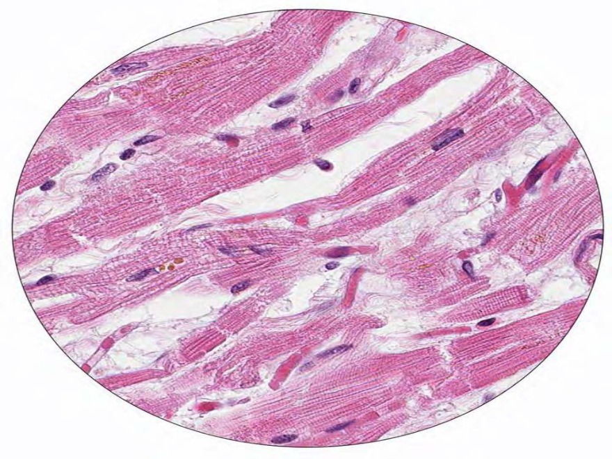
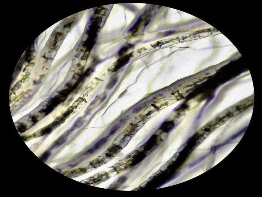
Identify the type of muscle tissue shown in the image below. 217
A) Skeletal muscle
B) Visceral smooth muscle
C) Cardiac muscle
D) Vascular smooth muscle
Correct Answer:C
Explanation:
The above image shows a longitudinal section of the cardiac muscle stained by hematoxylin and eosin.
On microscopy, cardiac muscle can be identified by:
Presence of striations similar to the skeletal muscle.
Presence of single and centrally placed nucleus. A perinuclear halo may be seen surrounding the nucleus.
The junctions between adjoining cardiac myocytes are seen as dark staining transverse lines running across the muscle fibre. These lines are called intercalated discs.
Cells within one fibre branch and join the cells in adjacent fibres.
Which of the following muscle fibers consists of spindle-shaped myocytes?
Image not available for this question yet.
A) Skeletal muscle
B) Smooth muscle
C) Cardiac muscle
D) Striated muscle
Correct Answer:B
Explanation:
Smooth muscles consist of spindle-shaped myocytes.
Smooth muscles are specialised for slow steady contraction under the influence of autonomic nerves and various hormones. They are also called as visceral muscles.
They are the major component of blood vessels, digestive, respiratory, urinary and reproductive tracts.
In microscopy, smooth muscles can be identified by:
In longitudinal section, the spindle-shaped muscle cells are seen. They are elongated, tapering and unstriated cells with a centrally placed elongated nucleus.
In transverse section, the spindle-shaped cells are cut at different places along its length and hence are seen as a group of cells with different shapes and sizes. The nucleus is seen only in those cells, which are cut through the centre.
Q1418.
Anatomy
Medium
4m
Image missing
Topic: Nervous and Endocrine SystemsSource: Internal
Explanation ready
Which of the following constitutes neurites of the neurons?
Image not available for this question yet.
A) Neurofibrils and dendrites
B) Dendrites and axons
C) Neurofibrils and axons
D) Neurofibrils, dendrites and axons
Correct Answer:B
Explanation:
The neurites of the neurons constitute the dendrites and the axon.
Neurites or neuronal processes refer to any projection arising from the cell body (soma) of a neuron. They are of two types:
Axon - a single, long projection arising from the cell body that carries the impulse away from the cell body. The axon ends by dividing into several branches called axon terminals, which synapses with the dendrites of another neuron. An axon may also end in relation to an effector organ such as muscle or a gland. It is free of Nissl granules.
Dendrites - multiple short projections arising from the cell body, that carry the nerve impulse towards the neuron. Nissl granules are present here.
The neuronal cytoskeleton consists of neurofilaments and microtubules. These are abundantly seen in the cell body, dendrites and the axon. The neurofilaments are heteropolymers of proteins that give tensile strength to the axon. Bundles of neurofilaments constitute a neurofibril.
Microtubules assist in axonal transport.
Q1419.
Anatomy
Medium
4m
Image missing
Topic: Nervous and Endocrine SystemsSource: Internal
Explanation ready
What is the Nissl-free zone from where an axon arises from a neuron called?
Image not available for this question yet.
A) Initial segment
B) Mesaxon
C) Axon hillock
D) Nodes of Ranvier
Correct Answer:A
Explanation:
The Nissl-free zone of a neuron from where an axon arises is called an axon hillock.
Nissl bodies or granules are aggregates of polyribosomes and rough endoplasmic reticulum. They are present in the cell body of a neuron and in large dendrites. Under light microscopy, they appear as basophilic granules. The affinity towards the basic dyes is due to the presence of a large amount of RNA which is acidic in nature.
The image below shows a photomicrograph of bluish-coloured Nissl bodies (indicated by arrows) in the cytoplasm of motor neurons.
Option A: The initial segment refers to the proximal part of the axon, that lies just beyond the axon-hillock. Thus, an axon-hillock is part of the cell body while the initial segment is part of an axon.
Option B: Mesaxon refers to the area where the plasma membrane of the Schwann cell fuses with itself around the axon. During the formation of the myelin sheath, the axon invaginates the Schwann cell. The plasma membrane of the Schwann cell fuses with itself and continues to wrap
around the axon to form multiple layers of the plasma membrane called the myelin sheath. The cytoplasm and nucleus of the Schwann cell are pushed to the periphery. The thin layer of the cytoplasm of the Schwann cell surrounding the myelin sheath is called neurilemma.
Option D: Each Schwann cell myelinates only a short segment of the axon. Hence, along
the length of the axon, there are several Schwann cells myelinating it. The unmyelinated portions of the axon present in between myelinated segments are known as nodes of Ranvier.
Q1420.
Anatomy
Medium
4m
Image ready
Topic: Nervous and Endocrine SystemsSource: Internal
Explanation ready
A 23-year-old patient was referred for evaluation of a progressive left-hand weakness and pain involving his right medial elbow, forearm, wrist and thumb. Suspecting pure neuritic leprosy, a nerve biopsy was taken and the image of the histological section is given below. What is the stain used in this section? 230
A) Sudan ƒlack
B) „iemsa
C) …smium tetroxide
D) Silver stain
Correct Answer:C
Explanation:
The stain used in this section is osmium tetroxide.
Osmium tetroxide is reduced to a black substance by unsaturated fatty acids present in the myelin sheath of the nerve fibers and hence, appears black on the section.
The pure neuritic form is a variant of leprosy characterized by the isolated involvement of peripheral nerve trunks with the absence of skin lesions. Nerve biopsy is the gold standard method for the diagnosis of pure neuritic leprosy.
Q1421.
Anatomy
Medium
4m
Image ready
Topic: Nervous and Endocrine SystemsSource: Internal
Explanation ready
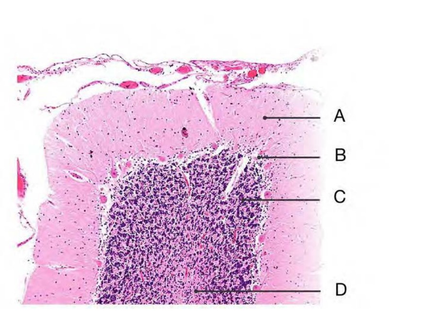
Match the marked structures to the correct layers of the cerebellar cortex.
A) A - 2, ƒ - †, ‡ - 3, D - ˆ
B) A - 2, ƒ - 3, ‡ - †, D - ˆ
C) A - 3, ƒ - 2, ‡ - †, D - ˆ
D) A - 3, ƒ - †, ‡ - 2, D - ˆ
Correct Answer:A
Explanation:
The structures marked in the given image are:
A - Molecular layer
B - Purkinje layer
C - Granular layer
D - White matter
The cerebellum is covered by pia mater which is seen as thin connective tissue, with blood vessels lying under it. The cerebellum consists of an outer grey matter (cerebellar cortex) and inner white matter.
Q1422.
Anatomy
Medium
4m
Image missing
Topic: Nervous and Endocrine SystemsSource: Internal
Explanation ready
232 In which layer of the cerebral cortex is the outer band of ƒaillarger seen?
Image not available for this question yet.
A) ‹xternal pyramidal layer
B) Internal granular layer
C) Internal pyramidal layer
D) ‹xternal granular layer
Correct Answer:B
Explanation:
The outer band of Baillarger is seen in the internal granular layer of cerebral cortex.
The cerebral cortex contains nerve fibers that run radially and tangentially. The tangential fibers are concentrated in the internal granular layer (layer IV) referred to as the outer band of Baillarger and in the ganglionic layer (layer V) referred to as the inner band of Baillarger. These bands are well developed in the sensory areas because of the concentration of terminal parts of the thalamocortical fibers. In the visual cortex, the outer band of Baillarger can be seen with the naked eye and is known as the stria of Gennari. It is because of this stria that the visual cortex is also referred to as the striate cortex.
The given image shows a micrograph of the visual cortex. The blue, horizontal band in the lower half of the image are the bands of Baillarger/the stria of Gennari. Subcortical white
matter (predominantly blue) is seen at the very bottom of the image.
Q1423.
Anatomy
Medium
4m
Image missing
Topic: Nervous and Endocrine SystemsSource: Internal
Explanation ready
An 8‘-year-old man was brought to the hospital with a history of a fall. Suspecting a skull bone fracture a lateral view ’-ray of the skull was taken which incidentally revealed a radio-opaque structure at the site of the pineal gland. Which of the following could be the most likely cause for the opacity?
Image not available for this question yet.
A) Žinealocytes
B) ‡orpora arenacea
C) Nervous conarii
D) ‡orpora amylacea
Correct Answer:A
Explanation:
The most likely cause for the opacity at the site of the pineal gland is corpora arenacea.
The pineal gland has a structure called the corpora arenacea (brain sand) located within the gland. These bodies are prone to an increase in calcification with advancing age. They are visible on x-ray and can be used as landmarks. Chemical analysis of the calcium deposits shows the different compositions, including calcium phosphate, magnesium phosphate, calcium carbonate, and ammonium phosphate.
Option A: The cells of the pineal gland are called pinealocytes that produces the hormone melatonin which works with the hypothalamus to regulate sleep-wake cycles.
Option D: Corpora amylacea are small eosinophilic inclusions frequently seen in histological sections of the brain, prostate, lung and uterus.
The image below is a histology section of the pineal gland showing dark stained corpora arenacea within the gland.
Q1424.
Anatomy
Medium
4m
Image ready
Topic: Nervous and Endocrine SystemsSource: Internal
Explanation ready
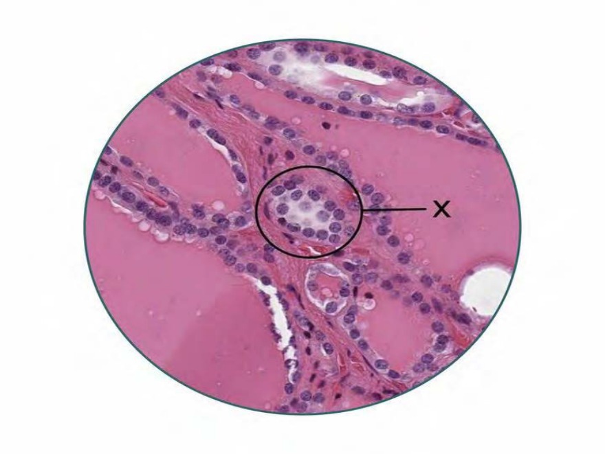
„iven below is a histological section of the thyroid gland. Identify the structure marked ’ in the image. 233
A) èollicular cells
B) ‡-cells
C) ‡olloid
D) çürthle cells
Correct Answer:D
Explanation:
The structure marked X is the C-cells or the parafollicular cells of the thyroid gland.
The C-cells are present between the follicular cells and the basement membrane or between the follicles. They secrete calcitonin.
The thyroid gland is covered by a fibrous capsule with septae dividing it into lobules. These lobules contain follicles lined by follicular cells. The shape of the follicular cells depends on the
activity of the gland. They're squamous in the inactive state, cuboidal to columnar in the active state. The cavity of the follicle contains colloid which appears pink in H and E staining.
Option D: Hürthle cells are oncocytes with eosinophilic granular cytoplasm and a vesicular nucleus with a large nucleolus found in the thyroid arising from follicular cells. They are seen in Hashimoto thyroiditis, Hürthle cell adenoma and Hürthle cell carcinoma of the thyroid.
Q1425.
Anatomy
Medium
4m
Image ready
Topic: Nervous and Endocrine SystemsSource: Internal
Explanation ready
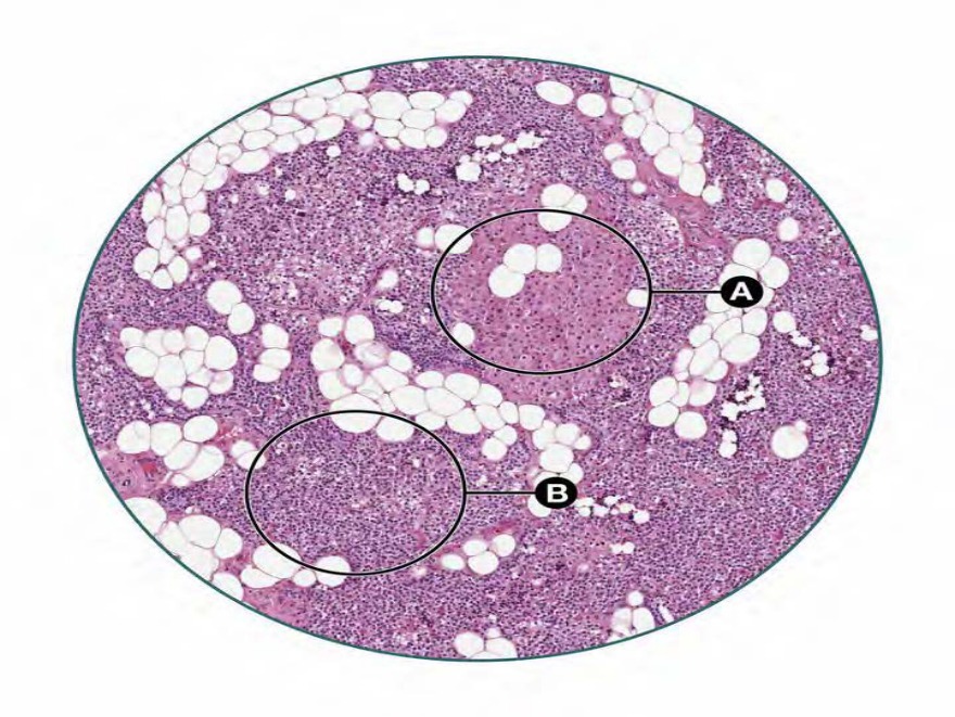
A 2†-year-old lady presented with a several-month-long history of abdominal pain, prolonged fatigue, bone, and joint pain. An MRI revealed a parathyroid gland tumour which was surgically resected. „iven below is a section of the parathyroid gland. Identify the structures marked A and ƒ. 234
A) A - follicular cells; ƒ - chief cells
B) A - …xyphil cells; ƒ - follicular cells
C) A - …xyphil cells; ƒ - ‡hief cells
D) A - ‡hief cells; ƒ - …xyphil cells
Correct Answer:C
Explanation:
The structures marked are A and B are oxyphil cells and chief cells respectively. The clinical history and the MRI findings are strongly suggestive of primary hyperparathyroidism secondary to a parathyroid adenoma.
The parathyroid gland has two cell types:
Chief cells (principal cells) - They are small round cells with vesicular nuclei, surrounded by a small amount of cytoplasm. They are numerous and produce parathormone.
Oxyphil cells - They are fewer and scattered between the chief cells. These are larger cells with
a smaller nucleus with eosinophilic cytoplasm (pink colored). Their function is not known
Primary hyperparathyroidism is usually due to a solitary parathyroid adenoma. The clinical signs associated with it can be remembered with the mnemonic - “bones, stones, groans, moans with fatigue overtones”.