Topic: Nervous and Endocrine SystemsSource: Internal
Explanation ready
In which of the following layers of adrenal cortex are the cells arranged in straight columns?
Image not available for this question yet.
A) •ona fasciculata
B) •ona glomerulosa
C) •ona reticularis
D) •ona medullaris
Correct Answer:A
Explanation:
The cells are arranged in straight columns in the zona fasciculata of the adrenal cortex. The adrenal cortex is divided into three layers. From outermost to deep, they are:
Zona glomerulosa - consists of small polyhedral cells arranged in inverted U-shaped formations or acinus-like groups. These cells produce mineralocorticoids
Zona fasciculata - consists of cells arranged in straight columns with sinusoids between the columns. These cells produce glucocorticoids.
Zona reticularis - It is so-called because it is made up of columns of cells that branch and anastomose with each other to form a kind of reticulum. These cells produce sex hormones -
progesterone, oestrogens and androgens.
Q1427.
Anatomy
Medium
4m
Image missing
Topic: Cardiovascular, Lymphatic and Respiratory SystemsSource: Internal
Explanation ready
Which of the following statements is true regarding the arteries?
Image not available for this question yet.
A) The fibrous elements present in the tunica intima and media run circularly
B) The fibrous elements of the tunica adventitia run longitudinally
C) Collagen fibers are predominantly present in the tunica media
D) Collagen fibers are predominantly present in the tunica intima
Correct Answer:B
Explanation:
The fibrous elements in the intima and the adventitia (mainly collagen) run longitudinally (i.e.,
along the length of the vessel), whereas those in the media (elastic tissue or muscle) run circularly.
The tunica adventitia consists of connective tissue in which collagen fibres are prominent. This layer prevents undue stretching or distension of the artery.
Structure of arterial wall:
The wall of an artery is made up of three layers, from inside out these include:
1. Tunica intima:
It is the innermost layer and consists of endothelium, which lies on the basal lamina.
Beneath the endothelium, there is a delicate layer of sub-endothelial connective tissue.
A membrane formed by elastic fibers surrounds the basal lamina. This membrane is called the internal elastic lamina.
2. Tunica media:
It consists of smooth muscle or elastic tissue, depending on which there are two types of
arteries:
muscular artery and elastic artery respectively.
The smooth muscle or elastic tissue is surrounded by a membrane made up of elastic fibers called, external elastic lamina.
3. Tunica adventitia:
It is the outermost layer and predominantly consists of connective tissue formed by collagen fibres.
This layer prevents the artery from being overstretched or excessively distended.
Q1428.
Anatomy
Medium
4m
Image missing
Topic: Cardiovascular, Lymphatic and Respiratory SystemsSource: Internal
Explanation ready
246 Which of the following statements is true regarding elastic arteries?
Image not available for this question yet.
A) They are also known as distributing arteries
B) The internal elastic lamina is distinct from the tunica media
C) Smooth muscles are absent in the tunica media
D) The elastic recoil helps in continuous flow of the blood
Correct Answer:A
Explanation:
The elastic recoil of the elastic arteries helps in continuous blood flow.
The large elastic arteries distend and recoil during systole and diastole respectively due to abundant elastic tissue. The recoiling acts as an additional force that pushes blood into smaller arteries. So, blood flows continuously through arteries but with some fluctuation in pressure during systole and diastole.
The main differences between the elastic and muscular arteries are shown in the table given below:
Structure Tunica intima
Tunica media
Tunica adventiti a
Elastic artery
The internal elastic lamina ca nnot be distinguished from t he surrounding tunica media
Predominantly of elastic fiber s.Few smooth muscle cells.
Relatively thin and consists mainly of elastic fibers
Muscular artery
The internal elastic lamina is distinct
Predominantly of smooth mu scle cells.Very few elastic fibe rs.
Relatively thicker and consist s of fibroblastic tissue.
Other options:
Option A: The elastic arteries are also known as conducting vessels, as their main function is to conduct the blood from the heart, whereas the muscular arteries are known as distributing vessels.
Option B: The internal elastic lamina in the elastic arteries is not distinct and cannot be differentiated from the tunica media as the structure of the internal elastic lamina is same as that of the elastic fibers present in tunica media of elastic arteries.
Option C: The tunica media of the elastic arteries is predominantly made up of elastic tissue. Between the elastic membranes, there is some loose connective tissue. Some smooth muscle cells may be present.
Q1429.
Anatomy
Medium
4m
Image missing
Topic: Cardiovascular, Lymphatic and Respiratory SystemsSource: Internal
Explanation ready
A researcher studying the COVID-19 virus recently discovered that anosmia is caused by the temporary loss of function of the sustentacular cells of the olfactory epithelium. Which of the following accurately describes these cells?
Image not available for this question yet.
A) Olfactory cells
B) Supporting cells
C) Receptor cells
D) Basal cells
Correct Answer:B
Explanation:
The sustentacular cells are supporting cells of the olfactory epithelium.
Their nuclei are oval and lie near the free surface of the epithelium. The free surface of each cell bears numerous microvilli (embedded in overlying mucous). The cytoplasm contains a yellow pigment (lipofuscin) that gives olfactory mucosa its yellow colour. In addition to their supporting function, sustentacular cells may be phagocytic, and the pigment in them may represent remnants of phagocytosed olfactory cells.
Q1430.
Anatomy
Medium
4m
Image missing
Topic: Cardiovascular, Lymphatic and Respiratory SystemsSource: Internal
Explanation ready
Given below is a histological section of the lung. Identify the structure marked X.
Image not available for this question yet.
A) Alveolus
B) Alveolar duct
C) Respiratory bronchiole
D) Terminal bronchiole
Correct Answer:B
Explanation:
The structure marked X in the above image is the lumen of the alveolar duct, which are tiny ducts that connect the respiratory bronchioles to alveolar sacs, each of which contains a collection of alveoli (small mucus-lined pouches made of flattened epithelial cells).
The coloured strips in the above image represent the location of different cells/structures in the trachea and bronchial tree.
On entering the lung, the principal bronchus divides into secondary, or lobar bronchi. Each lobar bronchus divides into tertiary, or segmental bronchi which further divides into the lobular bronchioles and then into terminal bronchioles.
Terminal bronchioles represent the most distal parts of the conducting passage. Each terminal bronchiole ends by dividing into respiratory bronchioles. Each respiratory bronchiole ends by dividing into a few alveolar ducts. Each alveolar duct ends in a passage, the atrium, which leads into a number of rounded alveolar sacs. Each alveolar sac is studded with a number of air sacs or alveoli.
A) Keratinised epithelium, submucosa, minor salivary glands
B) Keratinised epithelium, absent submucosa, no salivary glands
C) Non-keratinised epithelium, submucosa, minor salivary glands
D) Non-keratinised epithelium , absent submucosa, minor salivary glands
Correct Answer:A
Explanation:
The hard palate contains keratinised stratified squamous epithelium, has a submucosa, and contains minor salivary gland. This keratinised stratified squamous epithelium has regional variations and may be orthokeratinised or parakeratinised(found in hard palate and gingiva).
The bony structure of the hard palate is covered by a firmly attached mucosa in the central part. In the lateral parts, the hard palate also has a submucosal layer containing blood vessels. There are minor mucous type salivary glands in the submucosa in the posterior part of the hard palate.
(Note: In the orthokeratinized epithelium the cell nuclei disappear in the keratinized layer, whereas in the parakeratinized epithelium flattened, highly condensed nuclei remain in the cell cytoplasm of the keratinized layer until exfoliation)
A) Inner longitudinal and outer circular muscle fibres
B) Inner circular and outer longitudinal muscle fibres
C) only circular muscle fibres
D) only longitudinal muscle fibres
Correct Answer:B
Explanation:
The muscularis externa has an inner circular muscle fibers and outer longitudinal muscle fibers.
The gut wall receives its strength mainly from the muscularis externa, which is the thick layer of muscle that surrounds the submucosa. It is responsible for gut movements such as peristalsis.
Note:
Muscularis externa contains smooth muscle, except in upper part of esophagus where it has only striated muscle fibres.
The stomach has 3 layers in the muscularis externa, with an extra oblique layer.
In the colon, the outer longitudinal fibers join to form prominent bundles called the taenia
coli.
Given below is a histological section of the duodenum showing the inner and outer layers of muscularis externa.
A patient with suspected oesophagal carcinoma is planned for an endoscopic biopsy. A section of the lesion was taken and sent for histopathological examination. Which of the following layers of the gastrointestinal tract is not expected to be seen on microscopic examination of the section?
Image not available for this question yet.
A) Mucosa
B) Muscularis propria
C) Submucosa
D) Serosa
Correct Answer:D
Explanation:
The serosa is absent in the oesophagus and hence, is not expected to be seen on microscopic examination.
The oesophagus has an adventitia which is made up of dense fibrous tissue covering the muscularis externa. Only the lower part of the esophagus which is intra-abdominal has a layer of peritoneum covering it.
In general, the gut tube is made of the following layers (inner to outer side):
Mucosa - The inner-most layer is the mucous membrane with the following sub-layers:
Lining epithelium
Lamina propria - A layer of connective tissue supporting the lining epithelium
Muscularis mucosae - A thin layer of smooth muscle
Submucosa - Made of loose areolar tissue on which the mucous membrane rests
Muscularis externa - Thick layer of muscle around the submucosa (with inner circular and outer longitudinal fibres)
Which of the following statements is false about Paneth cells?
Image not available for this question yet.
A) They are rich in rough endoplasmic reticulum
B) Rich in zinc
C) Contain lysozyme
D) Foamy appearance
Correct Answer:D
Explanation:
The foamy appearance or frothy cytoplasm is not a feature of Paneth cells, but of Goblet cells. Mucus-secreting Goblet cells contain lipid or mucinous material with a foamy appearance.
Paneth cells (Enzyme cells) are present only in the deeper portions of the intestinal crypts. They contain many eosinophilic secretory granules. On microscopic examination, the Paneth cells are seen to contain a significant amount of rough endoplasmic reticulum. They are also rich in
zinc. The function of Paneth cells is to produce lysozyme that kills bacteria. They may also produce other enzymes.
A 42-year-old man presented to the clinic with a history of anemia, non-bloody diarrhoea and poor appetite. A diagnosis of celiac disease is suspected and a duodenal biopsy is taken for histopathological examination. Which of the following cells are not likely to be seen on microscopic examination of the section?
Image not available for this question yet.
A) Stem cells
B) Goblet cells
C) Paneth cells
D) Neck cells
Correct Answer:A
Explanation:
Neck cells are not likely to be seen on microscopy as they are not present in the small intestine. The neck cells are the mucous secreting cells that are present in the upper end (or ‘neck’) of the main gastric glands of the stomach.
Option A: Stem cells are the undifferentiated cells present in the wall of the intestinal crypts.
Option B: Goblet cells are mucous secreting cells. Each goblet cell has an expanded upper part that is distended with mucin granules. The nucleus is flattened and is situated near the base of the cell.
Option C: Paneth Cells (zymogen cells) are found only in the deeper parts of intestinal crypts. They contain prominent eosinophilic secretory granules.
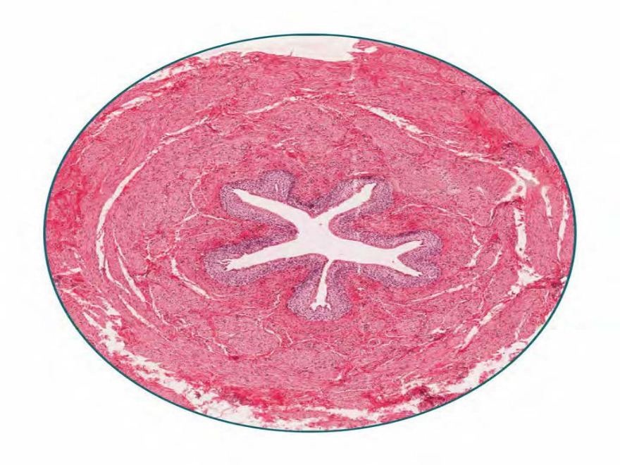
Which of the following structures does not form the portal triad of the liver?
Image not available for this question yet.
A) ‘epatic artery
B) ‘epatic vein
C) Bile duct
D) Portal vein
Correct Answer:B
Explanation:
Hepatic vein does not form part of the portal triad.
Portal canals are present at the angular intervals at the periphery of each hepatic lobule. They are filled with connective tissue and form a connective tissue network across the entire liver substance.
Each ‘canal’ consists of
A branch of the portal vein
A branch of the hepatic artery
An interlobular bile duct.
These three structures collectively form a portal triad.
The image given below shows a cross-section of the normal hepatic tissue showing the portal triad.
The transitional epithelium lines all of the following except
Image not available for this question yet.
A) Ureters
B) Minor calyx
C) Urinary bladder
D) Membranous urethra
Correct Answer:D
Explanation:
Transitional epithelium lines all the above structures except the membranous urethra.
Transitional epithelium is found in the renal pelvis and calyces, the ureter, the urinary bladder, and pre-prostatic amp; prostatic parts of the urethra. Because of this distribution, it is also called the urothelium.
Both in the male and female, the greater part of the urethra is lined by pseudostratified columnar epithelium. A short part adjoining the urinary bladder is lined by transitional epithelium, while the part near the external orifice is lined by stratified squamous epithelium.
The following are various parts of the urethra in males and their lining epithelium:
In which of the following structures is the type of epithelium as shown in the image seen?
A) Bile duct
B) Common bile duct
C) Skin
D) Urinary bladder
Correct Answer:D
Explanation:
The above image shows transitional epithelium and is seen in the urinary bladder.
The deepest layer has columnar cells, the middle layers are made up of polyhedral cells and the surface layer has cells that are large and shaped like an umbrella.
Transitional epithelium is found in the renal pelvis and calyces, the ureter, the urinary bladder, and part of the urethra. Because of this distribution, it is also called the urothelium. In the urinary bladder, it is usually observed that transitional epithelium can be stretched considerably without being damaged. When stretched it appears to be thinner and the cells become flattened or rounded.
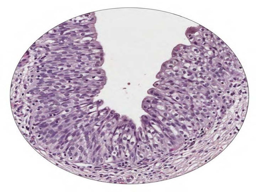
Which of the following are the epithelial supporting cells of the seminiferous tubules?
Image not available for this question yet.
A) Germ cells
B) Leydig cells
C) Sertoli cells
D) Peritubular cells
Correct Answer:C
Explanation:
Sertoli cells also called sustentacular cells are the epithelial supporting cells of the seminiferous tubules.
Sertoli cells are derived from the epithelial sex cords of the developing gonads. They are tall simple columnar cells. They surround the proliferating and differentiating germ cells forming pockets around these cells. Sertoli cells provide nutrients and phagocytose excess cytoplasm from the newly forming spermatozoa.
The given image is a section of seminiferous ducts showing the different types of cells.
Identify the structure shown in the histological image below. 267
A) Ureter
B) Epididymis
C) Seminal vesicle
D) Vas deferens
Correct Answer:D
Explanation:
Among the given options the histological section is most likely to be of the vas deferens or the ductus deferens.
The epithelium of this tube displays a pseudostratified columnar epithelium. It is surrounded by a prominent muscular layer. This layer contains inner and outer longitudinal muscle and a middle circular layer. An adventitia of connective tissue surrounds the muscularis layer. The ductus deferens extends from the epididymis to the ejaculatory ducts.
Q1443.
Anatomy
Medium
4m
Image missing
Topic: Skin Special Senses, Eye and EarSource: Internal
Explanation ready
Which of the following layers of epidermis is referred to as the prickle cell layer?
Image not available for this question yet.
A) Stratum basale
B) Stratum spinosum
C) Stratum granulosum
D) Stratum corneum
Correct Answer:B
Explanation:
Stratum spinosum of the epidermis is also referred to as the prickle cell layer.
Stratum spinosum consists of inter-cellular attachments called desmosomes. During the Hamp;E mounting, the cells shrink and retract at places other than their attachment at the
desmosomes. Thus, these desmosomes appear like spines on the cells on microscopy, and hence the cells are called as prickle cells.
Q1444.
Anatomy
Medium
4m
Image missing
Topic: Skin Special Senses, Eye and EarSource: Internal
Explanation ready
286 In which of the following layers of the epidermis are multiple layers of intertwining anucleate keratinocytes seen?
Image not available for this question yet.
A) Stratum lucidum
B) Stratum basale
C) Stratum granulosum
D) Stratum corneum
Correct Answer:D
Explanation:
The stratum corneum (horny layer) consists of multiple layers of anucleate keratinocytes that often intertwine resulting in a basketweave appearance.
The deepest layer, stratum basale, contains the stem cells which divide and start moving superficially. As cells move more superficially, they begin to lose their nuclei and become progressively dehydrated, flattened and intertwined. Traces of flattened nuclei may be seen in some cells of stratum lucidum.
Stratum corneum, because of its thickness, prevents entry of microbes from the environment into the body as well as preventing leakage of fluids from the body to the environment. This is called barrier function.
Q1445.
Anatomy
Medium
4m
Image missing
Topic: Skin Special Senses, Eye and EarSource: Internal
Explanation ready
A young patient presented with an itchy, rough rash with silvery scales along the extensor surface of her elbows, knees and scalp. Histopathological examination of the skin sample showed marked elongation and clubbing of the structure marked X in the given image. Identify the structure marked X. 287
Image not available for this question yet.
A) Reticular dermis
B) Hair follicle
C) Papillary dermis
D) Epidermal pegs
Correct Answer:D
Explanation:
The clinical history is suggestive of psoriasis and the structure marked X are the epidermal pegs or Rete ridges, which are downward projections of the epidermis in the intervals between the dermal papillae.
Histopathological examination of the skin in a case of psoriasis will show clubbing and elongation of the Rete ridges as shown in the given image.
Q1446.
Anatomy
Medium
4m
Image missing
Topic: Skin Special Senses, Eye and EarSource: Internal
Explanation ready
In which of the following layers of the skin are melanocytes present?
Image not available for this question yet.
A) Stratum corneum
B) Stratum granulosum
C) Stratum basale
D) Dermis
Correct Answer:C
Explanation:
Melanocytes are present in the stratum basale of the epidermis.
They are the neural crest derivatives that migrate to the stratum basale during the embryonic period. One melanocyte accumulates for every five or six basal keratinocytes.
Melanocytes synthesise the pigment, melanin. This is then transferred along the dendritic processes of the melanocyte in vesicles called melanosomes. The tips of these processes are then phagocytosed by the surrounding keratinocytes, which then take up the melanin.
The image below shows multiple melanocytes marked with arrows in the stratum basale of the epidermis.
Q1447.
Anatomy
Medium
4m
Image missing
Topic: Skin Special Senses, Eye and EarSource: Internal
Explanation ready
Which of the following cells act as slow-adapting touch receptors?
Image not available for this question yet.
A) Melanocytes
B) Keratinocytes
C) Langerhans cells
D) Merkel cells
Correct Answer:D
Explanation:
Merkel cells act as slow-adapting mechanoreceptors and are sensitive to light touch sensation.
They are present in the stratum basale of the highly sensitive skin like fingertips and bases of the hair follicle. The base of the Merkel cells is connected to the unmyelinated sensory fibres penetrating the basal lamina.
Q1448.
Anatomy
Medium
4m
Image missing
Topic: Skin Special Senses, Eye and EarSource: Internal
Explanation ready
Identify the structure marked X in the given histological image of the dermis.
Image not available for this question yet.
A) Merkel cell
B) Meissner’s corpuscles
C) Pacinian corpuscles
D) Ruffini’s corpuscles
Correct Answer:C
Explanation:
The structure marked X in the histological image of the dermis is the Pacinian corpuscle which appears as a large ( 1 mm), onion-like structure in the dermis and hypodermis.
It consists of concentric lamellae of flattened Schwann cells and collagen surrounding an unmyelinated axon. They are very rapidly adapting mechanoreceptors and specialised for sensing coarse touch, pressure (sustained touch) and vibration.
Q1449.
Anatomy
Medium
4m
Image missing
Topic: Skin Special Senses, Eye and EarSource: Internal
Explanation ready
A 28-year-old man presented to the dermatology OPD with complaints of patchy hair loss. A punch biopsy of the scalp was taken to assess the cause of the hair loss. Given below is the histological section of the hair follicle. Identify the structure marked Y in the image
Image not available for this question yet.
A) Cuticle
B) Medulla
C) Inner root sheath
D) Outer root sheath
Correct Answer:D
Explanation:
The structure marked Y in the above image is the outer root sheath. The hair is made up of the following layers (from within outwards):
Medulla - consisting of vacuolated and moderately keratinised cells.
Cortex - Consisting of densely packed and heavily keratinised cells.
Inner root sheath - This includes cuticle, inner Huxley's layer and outer Henle's layer.
Outer root sheath - These appear as clear cells as they contain glycogen.
Glassy membrane
Connective tissue sheath
Q1450.
Anatomy
Medium
4m
Image missing
Topic: Skin Special Senses, Eye and EarSource: Internal
Explanation ready
Which of the following accurately describes the arrector pili muscle?
Image not available for this question yet.
A) Skeletal muscles
B) Smooth muscles
C) Mixture of skeletal and smooth muscle, with dominant skeletal muscle
D) Mixture of skeletal and smooth muscle, with dominant smooth muscle
Correct Answer:B
Explanation:
Arrector pili muscles are smooth muscles.
They connect the undersurface of the hair follicles to the papillary dermis and are supplied by sympathetic nerves. Contraction of these muscles leads to the erection of hair, tiny bumps on the skin and thus produces a 'goose flesh’ appearance. The other effect of the contraction of arrector pili is the squeezing out of the secretions of the sebaceous glands into the hair follicle. They are absent in facial, axillary and pubic hairs, eyelashes, eyebrows and hairs around the nostrils and external auditory meatus.