Topic: Embryonic Phase of DevelopmentSource: Internal
Explanation ready
Formation of which of the following indicates the beginning of gastrulation?
Image not available for this question yet.
A) Notochord
B) Neural groove
C) Neural pit
D) Primitive streak
Correct Answer:D
Explanation:
The formation of the primitive streak from the epiblast is an indicator of the beginning of gastrulation.
By the 15th day of development, some epiblast cells lying along the central axis, near the tail end of the embryonic disc, begin to proliferate. These proliferated cells form an elevation called primitive streak that bulges into the amniotic cavity.
Gastrulation is the process that forms all the 3 germ layers namely, ectoderm, mesoderm, and endoderm in the embryo. It occurs during the 3rd week of gestation.
The image given below shows the formation of primitive streak.
Q1302.
Anatomy
Medium
4m
Image missing
Topic: Embryonic Phase of DevelopmentSource: Internal
Explanation ready
Which of the following statements regarding intraembryonic mesoderm is false?
Image not available for this question yet.
A) It is also known as secondary mesoderm
B) It Is formed from the epiblast cells
C) It spreads throughout the embryonic disc
D) Most of the body tissues are formed from mesoderm
Correct Answer:C
Explanation:
The intraembryonic mesoderm spreads throughout the embryonic disc, except in the region of the prechordal plate and cloacal membrane. In these two regions, the ectoderm is in direct contact with the endoderm.
The image given below shows the direct contact of intraembryonic mesoderm with the endoderm.
The intraembryonic mesoderm or secondary mesoderm is formed by the invagination and movement of epiblast cells between the newly formed endoderm and the epiblast.
In the later stages of development, the ectoderm and endoderm mostly persist as lining epithelium. Mesoderm forms most of the tissues of the body, including bone, cartilage, connective
tissue, muscles (skeletal, smooth, cardiac), blood, lymph, and cardiovascular organs.
Q1303.
Anatomy
Medium
4m
Image missing
Topic: Embryonic Phase of DevelopmentSource: Internal
Explanation ready
Neurulation refers to the process of the formation of:
Image not available for this question yet.
A) Primitive streak
B) Neural plate
C) Notochord
D) Neural tube
Correct Answer:D
Explanation:
Neurulation refers to the process of the formation of the neural tube from the neural plate. The neural tube gives rise to the central nervous system.
The image given below shows the steps of neurulation:
Q1304.
Anatomy
Medium
4m
Image ready
Topic: Embryonic Phase of DevelopmentSource: Internal
Explanation ready
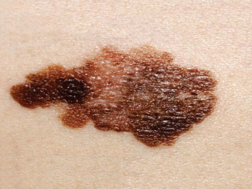
A 45-year-old woman presented with an itchy elevated lesion on her face as shown in the image. On dermoscopy, there is an atypical pigmented network. From which structure is the cell responsible for this lesion derived? 31
A) Lateral plate mesoderm
B) Intermediate mesoderm
C) Neural crest cell
D) Paraxial mesoderm
Correct Answer:A
Explanation:
The given image shows asymmetrical pigmented lesion on the face with an irregular border. This is suggestive of malignant melanoma arising from the melanocytes. Melanocytes are derived from neural crest cells (NCC).
During neurulation, when the neural folds elevate and fuse, the cells at the lateral border (the neural crest) of the neuroectoderm dissociate from their neighboring cells. These neural crest cells undergo an epithelial-to-mesenchymal transition while leaving the neuroectoderm and then migrate to the underlying mesoderm.
NCCs contribute to many organs and tissues and are sometimes referred to as the fourth germ layer:
In the trunk region, they form:
Melanocytes
Sensory ganglia
Sympathetic and enteric neurons
Schwann cells
Adrenal medulla cells
In the cranial region, they contribute to the:
Craniofacial skeleton
Neurons for cranial ganglia
Satellite glial cells
Melanocytes
Some of the other neural crest derivatives are:
C cells of the thyroid gland
Conotruncal septum in the heart
Odontoblasts
Smooth muscle cells to blood vessels to face and forebrain.
Option A: Lateral plate mesoderm is the lateral layer of the intraembryonic mesoderm. This is a thin layer. This layer develops a large cavity called the intraembryonic cavity and gives rise to a somatic or parietal mesodermal layer and a splanchnic or visceral mesodermal layer. The intraembryonic cavity gives rise to pericardial, pleural, and peritoneal cavities.
Option B: Intermediate mesoderm is a longitudinal strip of intraembryonic mesoderm that connects paraxial with the lateral plate mesoderm. The urinary and genital systems are derived
from the intermediate mesoderm.
Option D: Paraxial mesoderm is the thickened plate of the intraembryonic mesoderm close to the midline, on either side of the notochord. This forms the somitomeres and somites.
Q1305.
Anatomy
Medium
4m
Image missing
Topic: Embryonic Phase of DevelopmentSource: Internal
Explanation ready
Anomaly scan in a pregnant woman revealed renal agenesis in the fetus. Which of the following mesodermal layers is involved?
Image not available for this question yet.
A) Extra-embryonic mesoderm
B) Paraxial mesoderm
C) Intermediate mesoderm
D) Lateral plate mesoderm
Correct Answer:C
Explanation:
Kidneys are developed from the intermediate mesoderm.
After the notochord is formed, the intraembryonic mesoderm on either side can be seen organized into 3 layers:
Paraxial mesoderm - Thickened plate of the mesoderm close to the midline, on either side of the notochord. This forms the somitomeres and somites.
Intermediate mesoderm - Longitudinal strip of mesoderm that connects paraxial with the lateral plate mesoderm. The urinary and genital systems are derived from the intermediate mesoderm.
Lateral plate mesoderm - A thinner layer of mesoderm that lies laterally. This layer develops a large cavity called the intraembryonic coelom that gives rise to pericardial, pleural, and
peritoneal cavities.
Q1306.
Anatomy
Medium
4m
Image missing
Topic: Embryonic Phase of DevelopmentSource: Internal
Explanation ready
A 26-year-old man with congenital scoliosis is found to have hemivertebrae. The affected structures are derivatives of which layer of the mesoderm?
Image not available for this question yet.
A) Lateral mesoderm
B) Paraxial mesoderm
C) Intermediate mesoderm
D) Extraembryonic mesoderm
Correct Answer:B
Explanation:
The vertebrae are derived from somites which are derivatives of paraxial mesoderm. The paraxial mesoderm during development gets organized into segments called somitomeres and somites.
Somitomeres appear in the head region of the embryo. They are rounded structures and 7 in number. They form the mesoderm and muscles of the head and jaw.
From the occipital region downwards, somitomeres organize into somites. The first pair of somites arise in the occipital region of the embryo at approximately the 20th day of development.
From here, new somites appear in craniocaudal sequence at a rate of approximately 3 pairs per day.
By the end of the 5th week, 42-44 pairs of somites are present. There are 4 occipital, 8 cervical,
thoracic, 5 lumbar, 5 sacral, and 8 to 10 coccygeal pairs. The first occipital and the last five to seven coccygeal somites later disappear. The remaining somites form the axial skeleton.
Q1307.
Anatomy
Medium
4m
Image missing
Topic: Embryonic Phase of DevelopmentSource: Internal
Explanation ready
Which of the following gives rise to the pericardial cavity?
Image not available for this question yet.
A) Paraxial mesoderm
B) Intermediate mesoderm
C) Lateral plate mesoderm
D) Extraembryonic mesoderm
Correct Answer:C
Explanation:
The pericardial cavity is formed by the intraembryonic celom in the lateral plate mesoderm.
Within the lateral plate mesoderm, a large cavity known as intraembryonic celom is formed. The intraembryonic celom forms the pericardial, pleural, and peritoneal cavities.
The lateral plate mesoderm is divided by the intraembryonic celom to form:
Somatopleuric or parietal layer of intraembryonic mesoderm that is in contact with the ectoderm. This forms the parietal layer of peritoneal, pleural, and pericardial membranes.
Splanchnopleuric or visceral layer of intraembryonic mesoderm that is in contact with endoderm. This forms the visceral layer of the peritoneal, pleural, and pericardial membranes.
The image given below shows the somatopleuric and splanchnopleuric layers and the intraembryonic celom.
Q1308.
Anatomy
Medium
4m
Image missing
Topic: Embryonic Phase of DevelopmentSource: Internal
Explanation ready
Which of following structures becomes the primitive gut?
Image not available for this question yet.
A) Part of amniotic cavity lined by the mesoderm
B) Part of amniotic cavity lined by the endoderm
C) Part of yolk sac lined by the endoderm
D) Part of yolk sac lined by the mesoderm
Correct Answer:C
Explanation:
The primitive gut is derived from the part of the yolk sac lined by the endoderm.
With the development of the lateral folds of the embryo, a part of the yolk sac gets enclosed within the embryo, to form a tube-like structure. This structure is lined by the endoderm and is called the primitive gut.
Initially, the primitive gut has a wide communication with the yolk sac. Later, three different sections of the primitive gut can be recognized:
Foregut - Part of the primitive gut cranial to the communication with the yolk sac
Midgut - Middle part of the primitive gut in communication with the yolk sac
Hindgut - Part of the primitive gut caudal to the communication with the yolk sac
The image given below shows the sections of primitive gut.
Q1309.
Anatomy
Medium
4m
Image ready
Topic: Embryonic Phase of DevelopmentSource: Internal
Explanation ready
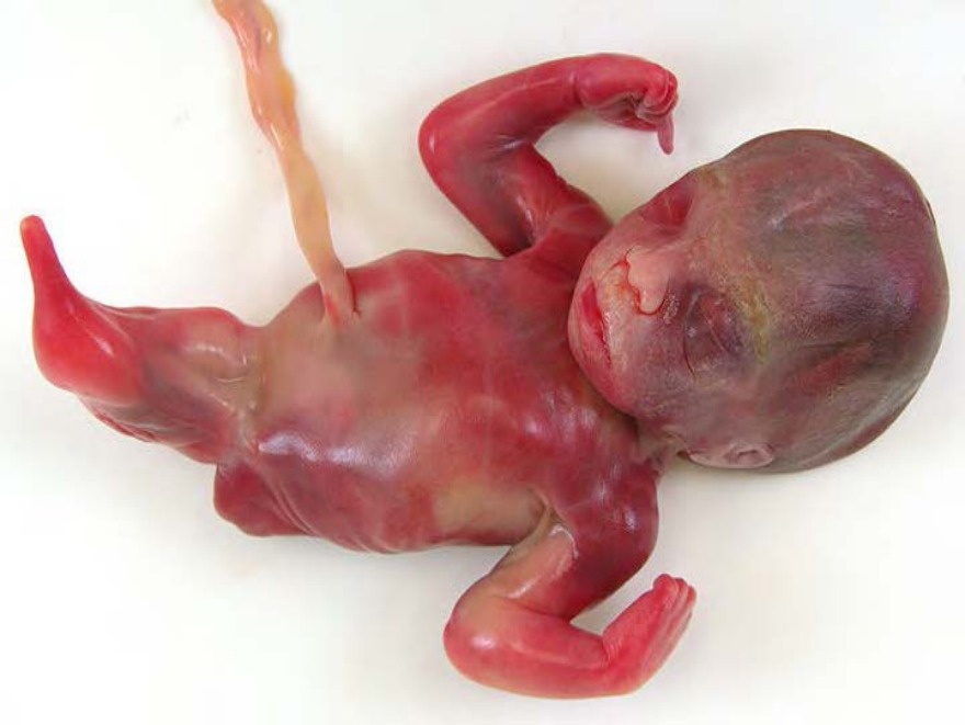
A 32-year-old primigravida who is a known diabetic, had a spontaneous miscarriage. The baby had the deformity as shown in the image below. Which of the following defects during embryonal development is likely to cause the given condition?
A) Insufficient formation of mesoderm
B) Persistence of pluripotent cells in primitive streak
C) Defect in the development of notochord
D) Loss of cells in the anterior midline of the germ disc
Correct Answer:A
Explanation:
The condition shown in the above image is sirenomelia. It is caused due to insufficient formation of mesoderm in the caudal most region of the embryo.
The mesoderm in the caudal region is involved in the formation of lower limbs, urogenital system, and lumbosacral vertebrae. Therefore, the defects seen in this condition include hypoplasia and fusion of the lower limbs, vertebral abnormalities, renal agenesis, imperforate anus, and anomalies of the genital organs.
During the third week of development, a process called gastrulation occurs which leads to the formation of all three germ layers. The process begins with the formation of the primitive streak, which develops into the primitive node and primitive pit. The primitive pit further invaginates and organizes itself into cells arranged in three layers, which eventually develop into ectoderm, mesoderm, and endoderm.
Gastrulation related abnormalities:
Sacrococcygeal teratomas occur due to remnants of pluripotent cells from the primitive streak.
They are the most common tumor in newborns, occurring at a frequency of 1 in 37,000 births. Sacrococcygeal teratomas contain tissues derived from all three germ layers.
When pluripotent cells from the primitive streak remain in the sacrococcygeal region, they proliferate to form sacrococcygeal teratomas.
Holoprosencephaly is a deficiency in the midline craniofacial structures. It occurs due to teratogenic exposure (such as high alcohol dose) during gastrulation which kills the cells in the anterior midline of the embryonic disc. In holoprosencephaly, the forebrain is small, the two lateral ventricles are often merged into a single ventricle, and the eyes are close together (hypotelorism).
Q1310.
Anatomy
Medium
4m
Image missing
Topic: Embryonic Phase of DevelopmentSource: Internal
Explanation ready
Which of the following blood vessels in the connecting stalk disappears during development?
Image not available for this question yet.
A) Right artery
B) Left artery
C) Right vein
D) Left vein
Correct Answer:C
Explanation:
The right vein in the connecting stalk disappears during development.
At first, the connecting stalk is at the caudal end of the embryonic disc. With the formation of the tail fold, the connecting stalk moves to the ventral part of the embryo, near the area of the umbilical opening.
The blood vessels connecting the placenta to the embryo pass through the connecting
stalk. Initially, there are 2 arteries and 2 veins. Later the right vein disappears (the left vein is left behind). The connecting stalk then consists of 2 arteries and 1 vein.
Q1311.
Anatomy
Medium
4m
Image missing
Topic: Embryonic Phase of DevelopmentSource: Internal
Explanation ready
Which of the following structures develops into Wharton’s jelly?
Image not available for this question yet.
A) Somatopleuric mesoderm
B) Splanchnopleuric mesoderm
C) Extraembryonic mesoderm
D) Paraxial mesoderm
Correct Answer:C
Explanation:
The extraembryonic mesoderm of the connecting stalk develops into a gelatinous substance known as Wharton’s jelly.
When the tail fold of the embryo is formed, the connecting stalk moves towards the ventral aspect of the embryo. The connecting stalk is now attached only in the region of the umbilical opening. The extraembryonic mesoderm of the connecting stalk becomes Wharton's jelly in the umbilical cord. Wharton's jelly protects the blood vessels in the umbilical cord.
Q1312.
Anatomy
Medium
4m
Image ready
Topic: Placenta, Fetal Membranes and TwinningSource: Internal
Explanation ready
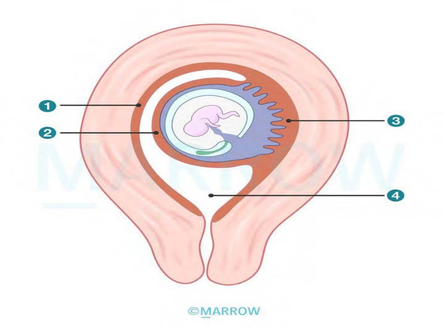
Identify the structures labeled in the image from 1 to 4.
A) Decidua basalis, Decidua capsularis, Decidua parietalis, Amniotic cavity
B) Decidua parietalis, Decidua capsularis, Decidua basalis, Amniotic cavity
C) Decidua basalis, Decidua capsularis, Decidua parietalis, Uterine cavity
The structures labeled in the above image are (1) Decidua parietalis, (2) Decidua capsularis, (3)
Decidua basalis, and (4) Uterine cavity.
The decidua is the modified endometrium formed under the influence of progesterone and human chorionic gonadotropin (hCG) secreted by the cells of syncytiotrophoblast. After the implantation of the blastocyst, the decidua gets divided into 3 parts as follows:
Decidua capsularis: part surrounding the embryo and separating it from the uterine cavity
Decidua basalis: part deeper to the developing blastocyst that contributes to the maternal component of the placenta
Decidua parietalis: part lining the rest of the uterine cavity
The image given below shows the parts of decidua.
Q1313.
Anatomy
Medium
4m
Image missing
Topic: Placenta, Fetal Membranes and TwinningSource: Internal
Explanation ready
Which of the following statements regarding chorionic villi is false?
Image not available for this question yet.
A) Primary villi contain cytotrophoblast and syncytiotrophoblast
B) Secondary villi contain extraembryonic mesoderm
C) Tertiary villi contain extraembryonic mesoderm
D) Secondary villi contain fetal capillaries
Correct Answer:D
Explanation:
Secondary villi do not contain fetal capillaries. Fetal capillaries are present in tertiary villi. Chorionic villi pass through the following stages during development:
Q1314.
Anatomy
Medium
4m
Image missing
Topic: Placenta, Fetal Membranes and TwinningSource: Internal
Explanation ready
Which of the following are macrophages present in the mesoderm of the chorionic villus?
Image not available for this question yet.
A) Langhans cells
B) Nitabuch’s cells
C) Hofbauer cells
D) Rohr's cells
Correct Answer:C
Explanation:
Hofbauer cells are fetal-derived macrophages present in the placenta.
Hofbauer cells are large (10–30 µm), pleomorphic cells, with highly vacuolated and granular cytoplasm. They are seen in the fetal villi of the placenta from the first trimester of pregnancy until birth.
The role of Hofbauer cells is not established yet, but they are thought to play a gate-keeper role in preventing the transmission of pathogens from the mother to the fetus.
Q1315.
Anatomy
Medium
4m
Image ready
Topic: Placenta, Fetal Membranes and TwinningSource: Internal
Explanation ready
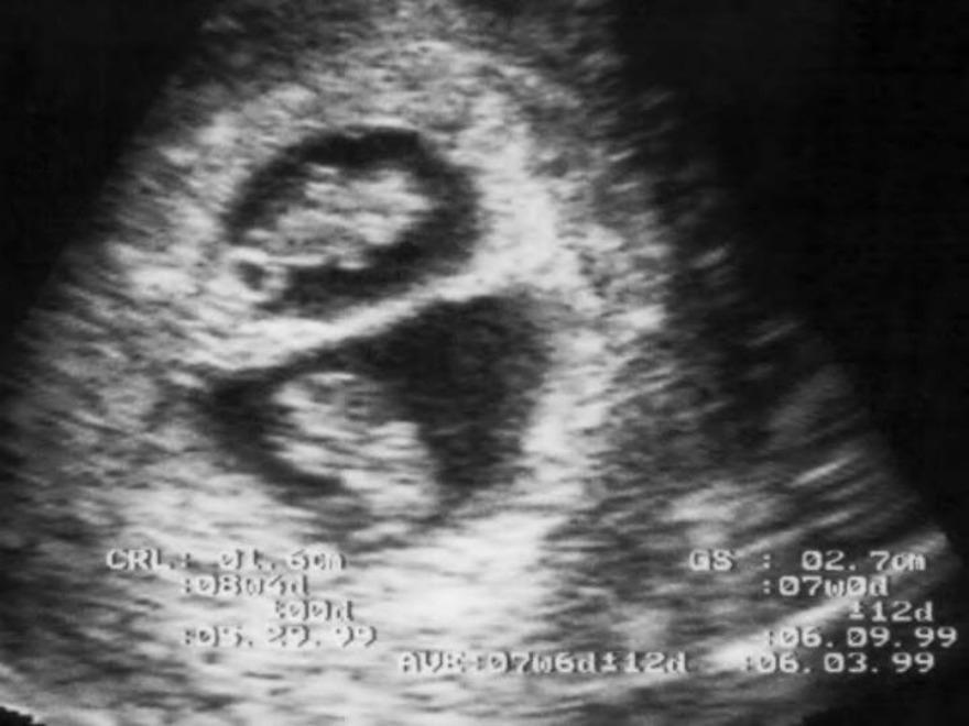
A pregnant lady comes for a routine antenatal checkup during the first trimester. USG examination reveals the following finding. Identify the type of twins in this patient.
A) Dichorionic, monoamniotic
B) Dichorionic, Diamniotic
C) Monochorionic, Diamniotic
D) Monochorionic, Monoamniotic
Correct Answer:B
Explanation:
The given scenario is suggestive of dichorionic, diamniotic twins. The image shows two separate amniotic cavities (diamniotic) and lambda sign (dichorionic).
In dichorionic twins, there is a thick triangular projection of placental tissue between the two gestational sacs. This is known as the lambda sign or twin peak sign. The following image shows the lambda/twin peak.
Dichorial, diamniotic twins can occur in 2 instances:
Dizygotic twins: The two fertilized ova get implanted at two separate sites and develop separate chorion and two separate amniotic cavities
Monozygotic twins: With early blastomere separation (up to 3rd day after fertilization), the two embryos formed will have separate chorionic and amniotic sacs, similar to dizygotic twins.
Q1316.
Anatomy
Medium
4m
Image ready
Topic: Placenta, Fetal Membranes and TwinningSource: Internal
Explanation ready
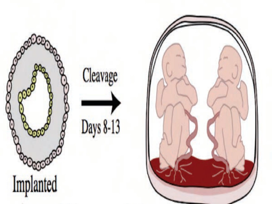
Identify the type of twin babies shown in the image below.
A) Di‹ygotic
B) Mono‹ygotic, Bichorial, Biamniotic
C) Mono‹ygotic, Monochorial, Biamniotic
D) Mono‹ygotic, Monochorial, Monoamniotic
Correct Answer:D
Explanation:
The image shows monozygotic, monochorial, monoamniotic twins. It depicts that a single zygote has separated between 8-13 days and therefore resulted in twins implanted at one site,
having the same chorion and having a single amniotic cavity.
The monozygotic, monochorionic, monoamniotic twins develop due to the duplication of the embryonic disc after the amniotic sac is already formed, i.e., between the 8th and 12th day after fertilization.
The core of the branchial arch is derived from which of the following?
Image not available for this question yet.
A) Ectoderm and mesoderm
B) Endoderm and neural crest cells
C) Mesoderm and neural crest cells
D) Ectoderm and endoderm
Correct Answer:C
Explanation:
The core of each pharyngeal arch (branchial arch) is derived from mesoderm and neural crest cells.
Pharyngeal arches initially have identical structures:
Surface ectoderm on the outside
A mesenchymal core derived from mesoderm
Endoderm derived epithelium on the inside
The mesoderm in the arches is derived from paraxial mesoderm and lateral plate mesoderm. This is invaded by the neural crest cells that contribute to the formation of skeletal elements and connective tissue of the head and neck region.
Which of the following pharyngeal arches disappear soon after formation?
Image not available for this question yet.
A) 3rd Arch
B) 4th Arch
C) 5th Arch
D) 6th Arch
Correct Answer:C
Explanation:
The 5th pharyngeal arch disappears soon after its formation, so only 5 arches remain.
Pharyngeal arches appear in the 4th or 5th week of development and contribute to the formation of the head and neck region. They are formed as a series of 6 thickenings in the cranial most part of the foregut (the future pharynx). They are craniocaudally numbered from I to VI. The 5th pharyngeal arch disappears, so only 5 arches remain.
The first pharyngeal arch is also known as the mandibular arch
The second pharyngeal arch is also known as the hyoid arch
The 3rd, 4th, and 6th pharyngeal arches have no special names
The site in the pharyngeal arch where the ectodermal cleft meets the endodermal pouch is known as:
Image not available for this question yet.
A) Pharyngeal cleft
B) Pharyngeal pouch
C) Pharyngeal membrane
D) Cervical sinus
Correct Answer:C
Explanation:
The pharyngeal membrane is the location where the ectoderm of the pharyngeal cleft meets the endoderm of the pharyngeal pouch.
In the interval between any two adjoining pharyngeal arches, the endoderm extends outward in the form of a pouch (pharyngeal pouch) to meet the ectoderm, which dips into this interval as a pharyngeal cleft. The membrane formed where the pharyngeal cleft comes in contact with the pouch is called the pharyngeal membrane.
Initially, there are four pharyngeal membranes between the five arches. Only the first pharyngeal membrane develops and contributes to the formation of the tympanic membrane. The 2nd, 3th, and 4th pharyngeal membranes do not develop further into any structure. They get obliterated when the mesoderm from the 2nd arch overgrows and fills the succeeding clefts.
Which of the following structures is not derived from Meckel's cartilage?
Image not available for this question yet.
A) Anterior ligament of malleus
B) Spine of sphenoid
C) Stapes
D) Sphenomandibular ligament
Correct Answer:C
Explanation:
The stapes is not derived from the Meckel's cartilage.
The cartilage of the first arch is Meckel's cartilage. Structures derived from it include:
Malleus
Incus
Anterior ligament of malleus
Spine of sphenoid
Sphenomandibular ligament
Note:
The mandible, maxilla, zygomatic bone, and part of the temporal bone are formed from the mesenchyme of the first arch and not from Meckel's cartilage.
The portion of Meckel’s cartilage that extends from the mandibular symphysis to mental foramen is probably incorporated into the intramembranous mandibular bone.
A 49-year-old woman came with complaints of dull aching pain in the throat and right side of the face. She also frequently gets pain while swallowing associated with earache and headaches. On CT scan, a calcified stylohyoid ligament was seen. This ligament is derived from:
Image not available for this question yet.
A) First pharyngeal arch
B) Second pharyngeal arch
C) Third pharyngeal arch
D) Fourth pharyngeal arch
Correct Answer:B
Explanation:
The stylohyoid ligament is derived from the cartilage of the second pharyngeal arch which is Reichert's cartilage.
Reichert's cartilage contributes to the development of the following structures: The dorsal portion forms:
Stapes
Styloid process of the temporal bone
Stylohyoid ligament
The ventral portion forms:
• The upper part of the body and the lesser cornu of the hyoid bone.
Note: This condition is called Eagle syndrome. It is a condition associated with the elongation of the styloid process or calcification of the stylohyoid ligament.
Which of the following muscles is affected in patients with mandibular nerve injury?
Image not available for this question yet.
A) Stylopharyngeus
B) Tensor tympani
C) Posterior belly of digastric
D) Levator veli palitini
Correct Answer:B
Explanation:
The tensor tympani muscle is affected in the mandibular nerve injury. The mandibular nerve supplies the muscle since it is derived from the first pharyngeal arch (mandibular arch).
Pharyngeal A rch
I
II
III IV
VI
Muscle derivatives
Muscles of mastication, mylo hyoid, anterior belly of digast ric, tensor tympani, and tens or veli palatini
Muscles of facial expression, stapedius, stylohyoid, platys ma, and posterior belly of dig astric
Stylopharyngeus
Larynx - cricothyroid; soft pa late - levator veli palatini; ph aryngeal constrictors
Which of the following is the nerve of 6th pharyngeal arch?
Image not available for this question yet.
A) Mandibular nerve
B) Superior laryngeal nerve of vagus
C) Glossopharyngeal nerve
D) Recurrent laryngeal nerve of vagus
Correct Answer:D
Explanation:
The recurrent laryngeal nerve of the vagus is the nerve of the 6th pharyngeal arch. The vagus nerve is distributed to the 4th and 6th arches as follows:
The tongue muscles are derived from the myotomes of the occipital somites.
The tongue muscles get their motor supply from the hypoglossal nerve (12th cranial nerve), except the palatoglossus muscle, which is supplied by the vagus nerve.
Development of the tongue
The anterior part of the tongue (body) is derived from the 1st pharyngeal arch. It is formed from
2 lateral lingual swellings and one medial swelling called the tuberculum impar.
The posterior part of the tongue (root) is derived from the mesoderm of the 2nd, 3rd, and part of the 4th arch. This part develops from a medial swelling called copula (hypobranchial eminence).
The posterior-most part of the tongue and the epiglottis are derived from a third swelling formed in the posterior part of the 4th arch.