A newborn had hypoplasia of the mandibles and zygomatic bones, malformed external ears, and slanted palpebral fissures. Abnormality in the development of which of the following pharyngeal arches will most likely lead to the given condition?
Image not available for this question yet.
A) Pharyngeal arch 1
B) Pharyngeal arch 2
C) Pharyngeal arch 3
D) Pharyngeal arch 6
Correct Answer:A
Explanation:
The given scenario is suggestive of Treacher Collins syndrome that results from abnormal development of the 1st pharyngeal arch (mandibular arch).
First arch syndromes are congenital defects caused due to the failure of the neural crest cells to migrate into the 1st pharyngeal arch. They are:
Craniofacial bone defects - Involves mandible (causing micrognathia), maxilla, and zygomatic arches
Ear defects - Involves the external ear, atresia of auditory canals, and abnormalities of the middle ear ossicles leading to bilateral conductive hearing loss
Eye defects - Down-slanting palpebral fissures and lower eyelid colobomas
Pierre Robin sequence (or Robin sequence) - Caused due to genetic defects or due to environmental factors. It can also be seen in oligohydramnios when the chin gets compressed against the chest in utero.
It is characterized by a triad of micrognathia, glossoptosis (posteriorly placed tongue), and
cleft palate
The primary defect is poor growth of the mandible (micrognathia)
As a consequence of this, the tongue is posteriorly placed (glossoptosis)
Additionally, the tongue remains in between the palatal shelves and they fail to fuse (cleft
During thyroidectomy, the surgeon accidentally damages a nerve. Post-op, the patient is not able to raise the pitch of his voice. Which arch is the affected structure derived from?
Image not available for this question yet.
A) 4th pharyngeal arch
B) 2nd pharyngeal arch
C) 3rd pharyngeal arch
D) 1st pharyngeal arch
Correct Answer:A
Explanation:
In the above scenario, the cricothyroid muscle is affected since it is responsible for high pitch phonation. It is derived from the 4th pharyngeal arch.
All the other intrinsic muscles of the larynx arise from the 6th pharyngeal arch.
Pharyngeal A rch
I
II
III IV
VI
Muscle derivatives
Muscles of mastication, mylo hyoid, anterior belly of digast ric, tensor tympani, and tens or veli palatini
Muscles of facial expression, stapedius, stylohyoid, platys ma, and posterior belly of dig astric
Stylopharyngeus
Larynx - cricothyroid; soft pa late - levator veli palatini; ph aryngeal constrictors
A 5-month-old infant was brought to the pediatric OPD for a routine checkup. The child had congenital hypoparathyroidism. CT scan revealed thyroid hypoplasia and absent thymus. Abnormality in the development of which of the following pharyngeal pouches will most likely cause the given features?
Image not available for this question yet.
A) Fourth and fifth pharyngeal pouch
B) First and second pharyngeal pouch
C) Second and third pharyngeal pouch
D) Third and fourth pharyngeal pouch
Correct Answer:D
Explanation:
The given clinical scenario of congenital hypoparathyroidism and thyroid hypoplasia, absent thymus on CT scan is suggestive of DiGeorge syndrome. The defect is most likely to be present in the development of third and fourth pharyngeal pouches.
Structures derived from various pharyngeal pouches (I-IV) are:
A 46-year-old woman presented with thyroid swelling. After evaluation, it was diagnosed as medullary thyroid cancer. Calcitonin produced from parafollicular C cells was used as a prognostic marker. These cells are derived from which structure?
Image not available for this question yet.
A) 3rd pharyngeal cleft
B) 3rd pharyngeal pouch
C) 4th pharyngeal cleft
D) Ultimobranchial body
Correct Answer:D
Explanation:
Calcitonin produced by parafollicular C-cells of the thyroid which is derived from the ultimobranchial body (4th pharyngeal pouch derivative).
The 4th pharyngeal pouch develops a small diverticulum medial to the main pouch called the ultimobranchial body. The ultimobranchial body is later incorporated into the thyroid gland and its cells give rise to the parafollicular or C-cells of the thyroid gland. These cells secrete a hormone called calcitonin that is involved in the regulation of the calcium level in the blood.
Clinical consideration: Both chromaffin cells of the adrenal medulla and parafollicular cells
(C-cells) of the thyroid originate from the neural crest cells. A common, germ-line mutation in neural crest cells can cause tumors in both the adrenal medulla and the thyroid, as seen in MEN types 2A and 2B.
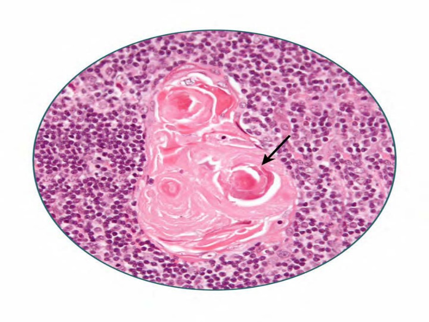
A patient diagnosed with thymoma underwent thymectomy. The histological section of the the organ is given below. The structure marked is derived from which pharyngeal pouch?
A) Fourth pharyngeal pouch
B) Third pharyngeal pouch
C) Second pharyngeal pouch
D) First pharyngeal pouch
Correct Answer:B
Explanation:
The image is showing Hassall’s corpuscles (thymic corpuscles) which are derived from the 3rd pharyngeal pouch.
The 3rd pouch gives rise to the thymus in the ventral part and the inferior parathyroid gland in the dorsal part. The thymus then moves away from the pharyngeal wall in a caudal and medial direction, pulling the inferior parathyroid with it. It reaches the anterior part of the thorax, where it fuses with its counterpart from the opposite side.
The endodermal cells of the thymus get invaded by the vascular mesoderm, which contains numerous lymphoblasts. Hassall’s corpuscles (thymic corpuscles) are derived from the endoderm of the 3rd pouch. The thymocytes are derived from bone and the cytoreticulum.
An 8-year-old girl was brought to the OPD with fever and throat pain for 3 days. On examination congestion and inflammation of the palatine tonsils were noticed. The affected structure is derived from which of the following?
Image not available for this question yet.
A) 1st pharyngeal pouch
B) 2nd pharyngeal pouch
C) 3rd pharyngeal pouch
D) Ultimobranchial body
Correct Answer:B
Explanation:
The diagnosis according to the given clinical vignette is acute tonsillitis affecting palatine tonsils. They are derived from the 2nd pharyngeal pouch.
The epithelium of the 2nd pharyngeal pouch proliferates to form solid cords known as tonsillar buds. The tonsillar buds penetrate into the surrounding mesoderm. These buds are then invaded by the mesodermal tissue to form the palatine tonsil. The capsule of the tonsil is formed by the condensed mesoderm. During the 3rd and 5th months, the tonsil is infiltrated by lymphatic tissue.
The intratonsillar cleft (tonsillar fossa) is the remnant of the 2nd pharyngeal pouch found in the adult.
The foramen cecum indicates the site of development of .
Image not available for this question yet.
A) Thyroid
B) Parathyroid
C) Thymus
D) Pituitary
Correct Answer:A
Explanation:
The foramen cecum indicates the site of development of thyroid.
The thyroid gland develops as a midline derivative of the pharynx between the 1st and
2nd pharyngeal pouches. Around the 5th week, a midline diverticulum is formed just caudal to the median tongue bud, at the site where foramen cecum is found later. This midline diverticulum passes caudally as the thyroglossal duct in front of the hyoid bone. The tip of this thyroglossal duct divides into two and their tissue mass proliferates to form the thyroid.
Tissues of the thyroid and their origin:
The epithelial tissue of the thyroid gland is derived from endoderm.
The connective tissue of capsule and interlobular septa are derived from the neural crest mesenchyme.
The parafollicular or C cells are derived from the ultimobranchial body, a small diverticulum of the 4th pharyngeal pouch.
Remnants of the thyroglossal duct can result in the formation of the thyroglossal cyst or fistula.
A 19-year-old girl presented with a painless swelling on the right side of the neck. CT-scan revealed a well-defined cystic mass at the angle of the mandible just in front of the anterior border of the sternocleidomastoid muscle. Persistence of which of the following structures during the development will most likely cause the given condition?
Image not available for this question yet.
A) First branchial cleft
B) Ultimobranchial body
C) Cervical sinus
D) Fourth branchial pouch
Correct Answer:C
Explanation:
The given scenario is suggestive of a branchial cyst that develops due to the persistence of the cervical sinus.
The cervical sinus is formed when the 2nd arch mesoderm overgrows the succeeding arches and overhangs them as shown in the image. The space between the overhanging 2nd pharyngeal
arch and the 3rd, 4th, and 6th arches is called the cervical sinus. The cavity of the cervical sinus is lined by ectoderm and normally gets obliterated.
In some individuals, a part of the cervical sinus may persist and give rise to swellings known as branchial cysts. It is present in the neck along the anterior border of the sternocleidomastoid muscle. These cysts are most commonly located just below the angle of the mandible. If the branchial cyst opens onto the surface, it becomes a branchial sinus.
The structures marked as “1” and “2” in the image below are developmentally derived from and , respectively. 69
Image not available for this question yet.
A) Notochord and sclerotome
B) Sclerotome and dermatome
C) Dermatome and notochord
D) Sclerotome and notochord
Correct Answer:D
Explanation:
The structure marked “1” is the outer annulus fibrosus and "2" is the inner nucleus pulposus of the intervertebral disc. They develop from the sclerotome and notochord, respectively.
The vertebral bodies and the annulus fibrosus (peripheral part of the intervertebral disc) are derived from the mesenchymal cells of the sclerotome.
The nucleus pulposus is a remnant of the notochord. The notochord regresses entirely in the region of vertebral bodies, but it persists in the central part of the intervertebral discs as
the nucleus pulposus.
Q636.
Anatomy
Medium
4m
Image missing
Topic: Cardiovascular and Respiratory SystemsSource: Internal
Explanation ready
Arrange the following structures of the primitive heart tube in cranial to caudal order.
Image not available for this question yet.
A) 2-3-4-1
B) 4-1-2-3
C) 1-4-2-3
D) 1-4-3-2
Correct Answer:D
Explanation:
The correct order of heart tube structures from cranial to caudal is bulbus cordis, primitive ventricle, primitive atrium, and the sinus venosus.
The 2 heart tubes formed in the cardiogenic area fuse, except in their caudal most end, to become an inverted Y-shaped structure. During this fusion, the central part of the heart tube expands to form the future outflow tract and ventricular regions. The caudal pole of the tube receives the venous drainage, while the cranial end pumps out blood into the arterial system.
Q637.
Anatomy
Medium
4m
Image missing
Topic: Cardiovascular and Respiratory SystemsSource: Internal
Explanation ready
A major part of the post-renal segment of the inferior vena cava develops from which of the following structures?
Image not available for this question yet.
A) Supracardinal vein
B) Umbilical vein
C) Anterior cardinal vein
D) Hepato-cardiac channel
Correct Answer:A
Explanation:
The post-renal segment of the inferior vena cava (IVC) mainly develops from the right supracardinal vein.
In embryonic life, the supracardinal veins get a large amount of venous drainage from the growing body wall. The right supracardinal vein persists and forms the greater part of the postrenal segment of the inferior vena cava.
The inferior vena cava is a composite vessel that develops from the remodeling of successive venous complexes. The veins contributing to the formation of IVC from upwards to below are as follows:
Cardiac termination of the right vitelline vein, also known as the right hepatocardiac channel
Anastomosis between right vitelline and right subcardinal veins (Hepatic segment of IVC)
Right subcardinal vein (Subcardinal or Pre-renal segment of IVC)
Anastomosis between right supracardinal and right subcardinal veins (Renal segment of IVC)
Part of the right supracardinal vein (Supracardinal or Post-renal segment of IVC)
Anastomosis between right supracardinal and right postcardinal veins
Part of the right posterior cardinal vein
Common iliac veins
Q638.
Anatomy
Medium
4m
Image missing
Topic: Cardiovascular and Respiratory SystemsSource: Internal
Explanation ready
Which of the following veins is not derived from the vitelline vein?
Image not available for this question yet.
A) Hepatic vein
B) Superior vena cava
C) Inferior vena cava
D) Superior mesenteric vein
Correct Answer:B
Explanation:
The superior vena cava is not derived from the vitelline vein.
Vitelline veins are developmental vessels passing between the yolk sac and the sinus venosus of the heart. Vitelline veins contribute to the development of the following structures:
Hepatic vein: Before entering the sinus venosus, the vitelline veins form a plexus around the duodenum and pass through the septum transversum. The liver cords grow into the septum breaking the course of this vein into an extensive vascular network known as hepatic sinusoids.
The sinusoids form the hepatic vein and drain into the inferior vena cava.
Portal vein: The anastomotic plexus formed by the vitelline veins around the duodenum develops into a single vessel called the portal vein.
Inferior vena cava: The hepatocardiac portion of IVC develops from the right hepatocardiac channel, which is derived from the right vitelline vein.
Superior mesenteric vein: This vein is derived from the right vitelline vein and drains the primary intestinal loop.
Q639.
Anatomy
Medium
4m
Image ready
Topic: Cardiovascular and Respiratory SystemsSource: Internal
Explanation ready
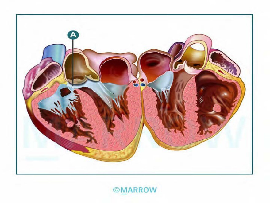
89 Marked structure is derived from the
A) Endocardial cushions
B) Septum primum
C) Left venous valve
D) Bulbous venosus
Correct Answer:A
Explanation:
The structure marked is the membranous part of the interventricular septum. It is derived from endocardial cushions.
Endocardial cushions are soft tissue masses that protrude in the atrioventricular and conotruncal regions.
Atrioventricular cushions are derived from overlying endocardial cells.
The conotruncal cushions are derived from the neural crest cells.
Endocardial cushions contribute to the formation of:
Atrial septa
Ventricular septa (membranous portion)
Atrioventricular canals
Atrioventricular valves
Aortic and pulmonary channels
Defects in endocardial cushion formation can result in cardiac malformations, including ASDs, VSDs, and defects involving the great vessels (transposition of the great vessels, common truncus arteriosus, and tetralogy of Fallot).
Q640.
Anatomy
Medium
4m
Image missing
Topic: Cardiovascular and Respiratory SystemsSource: Internal
Explanation ready
An infant with a continuous machine-like murmur is diagnosed with patent ductus arteriosus. When does the affected structure normally begin to close?
Image not available for this question yet.
A) 1-2 months
B) 1-2 weeks
C) 1-2 days
D) Immediately
Correct Answer:D
Explanation:
The ductus arteriosus gets functionally closed through contraction of its muscular wall almost immediately after birth. However, anatomical closure occurs only 2-3 weeks after birth.
In fetal life, prostaglandins relax the vascular smooth muscle and keep the ductus arteriosus open.
After birth, there is high oxygen content in the blood due to ventilation in the lung. This inhibits the production of prostaglandins. The ductus closes due to vascular smooth muscle contraction leaving a ligamentous remnant known as ligamentum arteriosum.
Note: Premature infants can be treated with prostaglandin synthesis inhibitors (such as indomethacin) to promote closure of the ductus arteriosus.
Q641.
Anatomy
Medium
4m
Image missing
Topic: Cardiovascular and Respiratory SystemsSource: Internal
Explanation ready
91 A newborn developed cyanosis soon after birth. Chest X-ray showed the characteristic egg-on-a-string appearance. Which of the following mechanisms during embryonic development will most likely lead to the given condition?
Image not available for this question yet.
A) Failure of cono-truncal ridge to fuse and descend
B) Aortico-pulmonary septum not following its spiral course
C) Anterior displacement of aortico-pulmonary septum
D) Non-fusion of aortico-pulmonary septum with truncus arteriosus
Correct Answer:B
Explanation:
The egg-on-a-string appearance on chest X-ray is characteristically seen in the transposition of great arteries (TGA). It is considered to arise due to abnormal spiraling of the aortopulmonary septum (conotruncal septum).
In this condition, the aortopulmonary septum fails to follow the normal spiral course and instead runs straight. This results in aorta arising from the right ventricle and the pulmonary trunk from the left ventricle. This condition is seen in 4.8/10,000 births.
Patent ductus arteriosus is commonly seen along with this condition. Sometimes, there is an associated defect in the membranous part of the interventricular septum.
Q642.
Anatomy
Medium
4m
Image missing
Topic: Cardiovascular and Respiratory SystemsSource: Internal
Explanation ready
Arrange the following phases of lung development and maturation in order, from the earlier phase to the later phase.
Image not available for this question yet.
A) 3-2-1-4
B) 2-1-4-3
C) 4-2-1-3
D) 2-4-3-1
Correct Answer:D
Explanation:
The correct order of phases of lung maturation is as follows: embryonic, pseudoglandular, canalicular, saccular phase.
The development and maturation of the lungs occur mainly in the following phases:
Phase
Embryonicphase (0–7 weeks)
Pseudoglandularphase (7–17 weeks)
Canalicularphase (17–27 wee ks)
Saccularphase/Alveolarphase
(28–32 weeks to after birth)
Developmental events
Appearance of lung buds and the main pulmonary arteries
Development of airways and blood vessels to the level of t he acinus
Formation of respiratory air ways and the development of the blood-air barrier
Development and secondary septation of alveoliAdjacent c apillary networks fuse to for m an extensive double capilla ry network
Q643.
Anatomy
Medium
4m
Image missing
Topic: Alimentary, Hepatobiliary systems, Pancreas and SpleenSource: Internal
Explanation ready
The mouth is derived from which of the following germ layers?
Image not available for this question yet.
A) Endoderm only
B) Endoderm and Mesoderm
C) Ectoderm and Endoderm
D) Ectoderm, Endoderm and Mesoderm
Correct Answer:C
Explanation:
The mouth is bidermal and derived partly from the stomatodeum (ectoderm) and partly from the cranial part of the foregut (endoderm).
The buccopharyngeal membrane, which is the demarcation between ectoderm and endoderm disappears by 4th week of development. The ectodermal and endodermal layers then become continuous with each other.
Q644.
Anatomy
Medium
4m
Image missing
Topic: Alimentary, Hepatobiliary systems, Pancreas and SpleenSource: Internal
Explanation ready
The primitive gut is derived from which of the following structures?
Image not available for this question yet.
A) Endoderm
B) Ectoderm
C) Amniotic cavity
D) Intraembryonic coelom
Correct Answer:A
Explanation:
The primitive gut is endodermal in origin.
When the cephalocaudal and lateral folding of the embryo occurs, a section of the endodermal germ layer gets enclosed within the embryo to form a tube-like structure called the primitive gut. The cephalic and caudal parts of the primitive gut end blindly are now known as the foregut and hindgut, respectively. The middle part is known as the midgut and is connected to the yolk sac through the vitelline duct.
Q645.
Anatomy
Medium
4m
Image missing
Topic: Alimentary, Hepatobiliary systems, Pancreas and SpleenSource: Internal
Explanation ready
Which of the following structures give rise to the duodenum?
Image not available for this question yet.
A) Foregut only
B) Midgut and Hindgut
C) Foregut and Midgut
D) Midgut only
Correct Answer:C
Explanation:
The duodenum develops from both the caudal part of the foregut and the cranial part of the midgut.
Structures originating from different sections of the primitive gut are as follows:
Foregut: pharynx, esophagus, stomach, duodenum (proximal), liver, gall bladder, pancreas
Midgut: duodenum (distal), small intestine (jejunum, ileum), appendix, cecum, ascending colon, proximal 2/3rd of transverse colon
Hindgut: distal 1/3rd of transverse colon, descending colon, proximal part of the rectum. From the cloaca (caudal part of hindgut) arise the distal part of the rectum and upper part of the anal canal.
Q646.
Anatomy
Medium
4m
Image missing
Topic: Alimentary, Hepatobiliary systems, Pancreas and SpleenSource: Internal
Explanation ready
The midgut loop normally herniates through the primitive umbilical ring into the extra-embryonic celom during the 6th-week of development. Failure of the intestinal loops to return to the abdominal cavity by 11th-week results in the formation of which of the following conditions in a baby?
Image not available for this question yet.
A) Omphalocele
B) Gastroschisis
C) Exstrophy of bladder
D) Ileal diverticulum
Correct Answer:A
Explanation:
Failure of the intestinal loops to return to the abdominal cavity during the 6th to 10th weeks results in omphalocele or exomphalos.
Omphalocele is a congenital malformation in which the abdominal viscera protrudes through a wide umbilical defect. The bowel loops are covered by amnion.
A small omphalocele contains only the intestines. A large omphalocele may contain other viscera such as the stomach, liver, spleen, and large intestine, along with the small intestine. Omphalocele is commonly associated with other congenital anomalies (e.g., trisomy 13, trisomy 18, or
Beckwith-Wiedemann syndrome) and causes increased levels of α-fetoprotein.
The images given below shows the omphalocele.
Note: Gastroschisis is the major disorder to consider in the differential diagnosis of an omphalocele. It can be differentiated from omphalocele by its absence of a membranous sac and paraumbilical location (commonly to the right of the umbilicus).
Q647.
Anatomy
Medium
4m
Image ready
Topic: Alimentary, Hepatobiliary systems, Pancreas and SpleenSource: Internal
Explanation ready
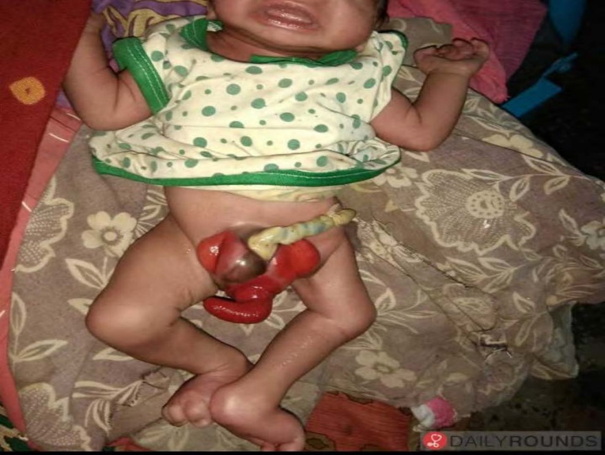
You note the given finding in a baby admitted to the pediatric ward. Which of the following defects in the intrauterine life is the cause of this condition?
A) Failure of the intestinal loops to return from umbilicus
B) Defective closure of body wall in the abdominal region
C) Defective closure of body wall in the pelvic region
D) Defective closure of the vitellointestinal duct
Correct Answer:C
Explanation:
The condition shown in the image is cloacal exstrophy, which is caused by the failure of the body wall to close in the pelvic region.
The given image shows the classic feature of cloacal exstrophy, which includes an exstrophic central bowel area flanked by two hemibladders:
Bladder exstrophy: The bladder is seen turned inside out (exstrophy means turned inside out)
and visible as 2 hemibladders on either side.
Bowel exstrophy: The central defect is the prolapsed intestinal loop that appears as the elephant trunk deformity. The central bowel field usually contains an ileocecal segment that has prolapsed.
In addition, omphalocele can be noted in the umbilical region in this image.
Exstrophy of the bladder or cloaca occurs due to abnormal closure of the body wall in the pelvic region:
Exstrophy of the bladder: The bladder mucosa is exposed to the surface. In male infants, epispadias is seen additionally.
Exstrophy of the cloaca: The closure of the ventral body wall is more severely affected than in bladder exstrophy. There is the involvement of the bladder and rectum, both of which are derived from the cloaca. In addition, there is defective development of the urorectal septum leading to anal canal malformations and imperforate anus. As the genital swellings are widely separated, external genitalia will be defective.
Cloacal exstrophy can present as part of OEIS syndrome, in which multiple anomalies are seen, where O stands for Omphalocele, E for Exstrophy of bladder or cloaca, I for Imperforate anus, and S for Skeletal anomalies.
Q648.
Anatomy
Medium
4m
Image missing
Topic: Alimentary, Hepatobiliary systems, Pancreas and SpleenSource: Internal
Explanation ready
The septum transversum develops from which of the following structures?
Image not available for this question yet.
A) Surface ectoderm
B) Mesoderm
C) Neuroectoderm
D) Endoderm
Correct Answer:B
Explanation:
Septum transversum develops from the mesoderm.
Septum transversum is a thick mass of mesodermal tissue that occupies the space between the developing thoracic cavity and yolk stalk in the early embryo. It works as a partition to separate thoracic and abdominal cavities.
The septum transversum gives rise to:
The central tendon of the diaphragm
The ventral mesentery of the foregut
The connective tissue of the liver
Q649.
Anatomy
Medium
4m
Image missing
Topic: Alimentary, Hepatobiliary systems, Pancreas and SpleenSource: Internal
Explanation ready
A 45-year-old man presented with chronic abdominal and back pain. CT scan of the abdomen was done, which incidentally revealed pancreatic divisum. Which of the following mechanisms during embryonic development leads to the given condition?
Image not available for this question yet.
A) Persistence of the proximal portion of dorsal pancreatic bud
B) Failure of fusion of the dorsal and ventral pancreatic buds
C) Failure of regression of dorsal pancreatic bud
D) Failure of regression of ventral pancreatic bud
Correct Answer:B
Explanation:
Failure of fusion of the dorsal and ventral pancreatic buds leads to a condition called pancreas divisum.
The developing pancreatic ducts usually fuse in such a way that most of the dorsal duct drains into the proximal part of the ventral duct. The proximal portion of the dorsal duct usually persists as an accessory duct. In about 10 of the population, the two ducts do not fuse, resulting in pancreatic divisum (pancreas divisum) where separate drainage into the duodenum is maintained.
The image given below shows the development of the pancreas.
The annular pancreas occurs when the right portion of the ventral bud migrates along its normal route, but the left migrates in the opposite direction. In this case, the duodenum is surrounded by pancreatic tissue.
Q650.
Anatomy
Medium
4m
Image ready
Topic: Face, Nose Palate, Eye, EarSource: Internal
Explanation ready
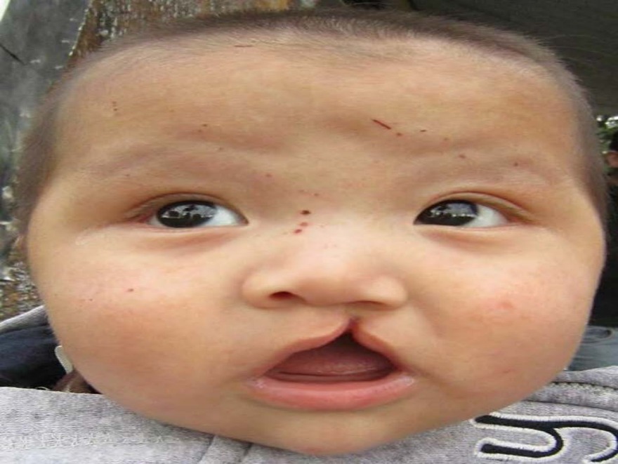
A 2-month-old baby was brought to the clinic with an anomaly as shown in the image. This occurs due to the failure of fusion between which structures? 132
A) Lateral nasal prominence and mandibular prominence
B) Lateral nasal prominence and maxillary prominence
C) Medial nasal prominence and mandibular prominence
D) Medial nasal prominence and maxillary prominence
Correct Answer:D
Explanation:
The image shows cleft lip which occurs when the maxillary prominences fail to fuse with the medial nasal prominences.
Various defects of fusion of prominences in the face region are as follows:
A cleft lip is due to the failure of the fusion of maxillary prominence with medial nasal prominence. It can be unilateral or bilateral. It can vary in severity from a small notch in the upper lip to a double cleft extending into both the nostrils. There is also associated underdevelopment of the part of the maxilla that bears the incisors.
Median (midline) cleft lip is a rare defect occurring due to failure of the two medial nasal prominences to fuse in the midline.
The oblique facial cleft is caused by the failure of the maxillary prominence to fuse with the lateral nasal prominence along the line of the nasolacrimal groove. The nasolacrimal duct is exposed to the surface in these cases.