Topic: Face, Nose Palate, Eye, EarSource: Internal
Explanation ready
The primary palate is formed from which structure?
Image not available for this question yet.
A) Medial nasal prominences
B) Lateral nasal prominences
C) Maxillary prominences
D) Frontonasal prominences
Correct Answer:A
Explanation:
The primary palate (premaxilla) develops from the intermaxillary segment that is formed by the fusion of the two medial nasal prominences.
The intermaxillary segment has the following components:
Labial component - forms the philtrum of the upper lip
Upper jaw component - forms the medial part of the maxillary bone with the four upper incisor teeth
Palatal component - forms the triangular area in the front called the primary palate (premaxilla)
Q652.
Anatomy
Medium
4m
Image missing
Topic: Face, Nose Palate, Eye, EarSource: Internal
Explanation ready
Arrange the following structures formed during the development of the eye in their order of appearance.
Image not available for this question yet.
A) 1-2-3-4
B) 2-4-3-1
C) 4-1-2-3
D) 1-3-4-2
Correct Answer:C
Explanation:
The order of the structures formed during eye development is Optic sulcus ° Optic vesicle °
Optic stalk ° Optic cup.
Q653.
Anatomy
Medium
4m
Image missing
Topic: Face, Nose Palate, Eye, EarSource: Internal
Explanation ready
The muscles that cause constriction and dilation of the pupil are derived from:
Image not available for this question yet.
A) Ectoderm
B) Endoderm
C) Mesoderm
D) Neural crest cells
Correct Answer:A
Explanation:
The musculature of the iris, including the sphincter pupillae and dilator pupillae, are derived from the ectoderm of the optic cup.
Most of the muscles of the body develop from mesoderm. The muscles which develop from ectoderm are:
Muscles of the iris (sphincter and dilator pupillae)
Arrectores pilorum of skin
Myoepithelial cells lining the ducts of sweat glands
Q654.
Anatomy
Medium
4m
Image missing
Topic: Face, Nose Palate, Eye, EarSource: Internal
Explanation ready
A newborn had complete absence of the external auricle and auditory meatus. There are no other physical deformities. Renal function is normal. Abnormalities in the development of which of the following structures during the embryonic development lead to the given condition?
Image not available for this question yet.
A) Third and fourth pharyngeal arches
B) Second and third pharyngeal arches
C) First and second pharyngeal arches
D) First and third pharyngeal arches
Correct Answer:C
Explanation:
Anotia (complete absence of the ear pinna) occurs due to the defect in the development of 1st and 2nd pharyngeal arches.
The pinna develops from 6 tissue elevations called the auricular hillocks formed around the margins of the dorsal portion of the 1st pharyngeal cleft.
3 hillocks are from the 1st pharyngeal (mandibular) arch
3 hillocks are from the 2nd pharyngeal (hyoid) arch.
The mandibular arch contributes to the tragus of the pinna only, while the rest of the pinna is derived from the hyoid arch. Invagination of the first pharyngeal arch gives rise to the external auditory meatus.
Previously, it was believed that the external auditory meatus develops from the first pharyngeal cleft. But recent studies suggest that the first pharyngeal arch gives rise to the external auditory meatus.
Q655.
Anatomy
Medium
4m
Image missing
Topic: Nervous System and Endocrine GlandsSource: Internal
Explanation ready
A 1-year-old baby with loss of pharyngeal reflex was diagnosed to have infantile progressive bulbar palsy. Which of the following gives rise to the affected structure?
Image not available for this question yet.
A) Prosencephalon
B) Mesencephalon
C) Rhombocephalon
D) None of the above
Correct Answer:C
Explanation:
In infantile progressive bulbar palsy, the affected structure is the medulla oblongata. It arises from the rhombocephalon (hindbrain).
The rhombencephalon is further subdivided into a cranial part (metencephalon) and a caudal part
(myelencephalon).
Metencephalon - forms the pons and cerebellum
Myelencephalon - forms the medulla oblongata
Q656.
Anatomy
Medium
4m
Image missing
Topic: Nervous System and Endocrine GlandsSource: Internal
Explanation ready
What is the flexure between the metencephalon and the myelencephalon called?
Image not available for this question yet.
A) Cephalic flexure
B) Mesencephalic flexure
C) Pontine flexure
D) Cerebellar flexure
Correct Answer:C
Explanation:
The flexure between the metencephalon and the myelencephalon is called as the pontine flexure.
The prosencephalon, mesencephalon, and rhombencephalon are initially arranged craniocaudally. Due to differential growth of different regions of the brain, various flexures form between them:
Note: A deep furrow called the rhombencephalic isthmus separates the mesencephalon from the metencephalon.
Q657.
Anatomy
Medium
4m
Image ready
Topic: Nervous System and Endocrine GlandsSource: Internal
Explanation ready
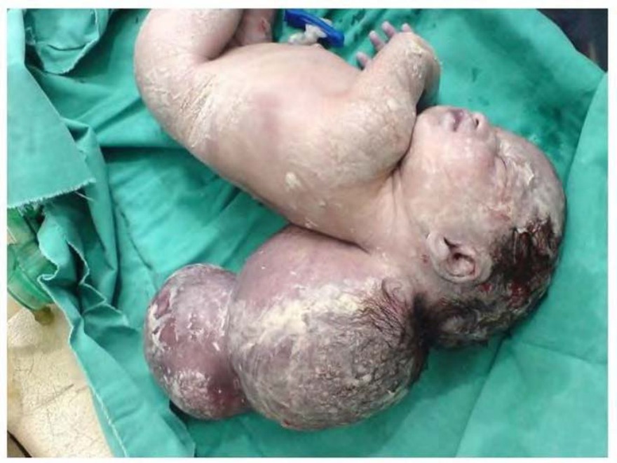
Identify the neural tube defect shown in the image.
A) Spina bifida
B) Rachischisis
C) Encephalocele
D) Myelocele
Correct Answer:C
Explanation:
The image shows a newborn with encephalocele.
Encephaloceles are caused due to defects in the skull through which the meninges and/or neural tissue herniate. The squamous part of the occipital bone is the most frequently affected bone. The bone may be partially or totally lacking.
Based on the degree of defect and tissues herniating, they are classified as follows:
Meningocele - Defect is small and only meninges bulge through it
Meningoencephalocele - Defect is large and a part of brain bulges with the meninges
Meningohydroencephalocele - Defect is large and a part of the brain and ventricle bulge with the meninges
Q658.
Anatomy
Medium
4m
Image ready
Topic: Nervous System and Endocrine GlandsSource: Internal
Explanation ready
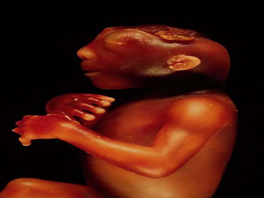
Identify the defect shown in the image. 149
A) Encephalocele
B) Spina bifida
C) Anencephaly
D) Meningoencephalocele
Correct Answer:C
Explanation:
The image shows the congenital defect of anencephaly.
Failure of the cranial end of the neural tube to close is known as exencephaly. As a result of this, the skull vault does not form. This leaves the malformed brain exposed.
The exposed brain tissue degenerates, leaving a mass of necrotic tissue. This defect with the absence of a major portion of the brain, skull, and scalp is known as anencephaly. The brain stem remains intact.
In some cases, the closure defect of the neural tube extends caudally, and the spinal cord is affected. This condition is known as craniorachischisis, where there is anencephaly plus a large defect involving the spine.
Q659.
Anatomy
Medium
4m
Image missing
Topic: Nervous System and Endocrine GlandsSource: Internal
Explanation ready
A ΍-year-old male patient underwent an MRI scan for the evaluation of stroke. Incidentally, it was found that the pineal gland was absent. Defect in the development of which of the following structures will likely lead to pineal agenesis?
Image not available for this question yet.
A) Telencephalon
B) Diencephalon
C) Metencephalon
D) Myelencephalon
Correct Answer:B
Explanation:
The pineal gland develops from the diencephalon. Hence, defect in its development can cause pineal agenesis.
The most caudal part of the roof plate of the diencephalon develops into the pineal gland. It is also known as the pineal body or epiphysis cerebri. The pineal body initially appears as an epithelial thickening in the midline. It begins to evaginate by the 7th week and becomes a solid organ.
The images given below show the pineal gland:
Q660.
Anatomy
Medium
4m
Image missing
Topic: Nervous System and Endocrine GlandsSource: Internal
Explanation ready
Which of the following zones of the adrenal cortex are present at birth?
Image not available for this question yet.
A) Žona fasciculata and Žona reticularis
B) Žona glomerulosa and Žona fasciculata
C) Žona glomerulosa and Žona reticularis
D) Žona glomerulosa, Žona fasciculata, and Žona reticularis
Correct Answer:B
Explanation:
The zona glomerulosa and zona fasciculata of the adult cortex is present at birth.
The differentiation of the cortical zones of the adrenal gland begins during the late fetal period.
Žona glomerulosa and zona fasciculata of the adult cortex are present at birth
Žona reticularis is formed during the 3rd year of life
Arrange the following stages of nephron development in order from the earlier stage to the later stage.
Image not available for this question yet.
A) 1-2-3-4
B) 4-3-2-1
C) 1-4-2-3
D) 1-4-3-2
Correct Answer:C
Explanation:
The correct order of the stages of development of the nephron from earlier to later is blastema stage --gt; vesicle stage --gt; comma 'C' stage --gt; S-shaped stage.
The tip of the ureteric bud, which is growing towards the metanephric blastema, becomes dilated to form an ampulla. The ampulla induces the cells of the metanephric blastema to develop into a nephron.
The cells of the metanephric blastema pass through the following stages when developing into a nephron:
The trigone on the posterior wall of the urinary bladder is formed by which structure?
Image not available for this question yet.
A) Mesonephric ducts
B) Metanephric blastema
C) Cloacal membrane
D) Urogenital sinus
Correct Answer:A
Explanation:
The trigone on the posterior wall of the urinary bladder is formed by the incorporation of the lower ends of the mesonephric ducts.
After this incorporation, the ureters now open separately into the bladder, near the opening of the mesonephric duct. Due to the ascent of the kidneys, the ureteral openings move farther cranially, while the openings of the mesonephric ducts move close together. The triangular area between these ducts (trigone of the bladder) is thought to arise from the terminal portion of the mesonephric ducts.
Recent update: It was considered that the epithelium of the trigone is initially mesodermal and is later replaced by the endoderm-derived transitional epithelium. However, current research indicates that there is no mesodermal epithelial contribution to the bladder at the trigone (Gray's anatomy, 41st edition).
A 6-year-old girl presents with a large abdominal mass just superior to the pubic symphysis. The mass is tender on palpation and fixed. During surgery, it is seen that the mass is fluid-filled and connected to the umbilicus superiorly and to the urinary bladder inferiorly. 165 What is the diagnosis?
Image not available for this question yet.
A) Vitelline cyst
B) Horseshoe kidney
C) Polycystic disease of the kidney
D) Urachal cyst
Correct Answer:D
Explanation:
A palpable fluid-filled mass that is connected to the umbilicus superiorly and urinary bladder inferiorly is diagnostic of the urachal cyst.
During embryonic development, the upper end of the urogenital sinus communicates with the allantois, which lies in the umbilical cord. The allantois normally regresses to form a fibrous cord
called the urachus. In the adult, the urachus is seen as the median umbilical ligament. The remnants of the urachus that may be seen after birth are:
166 A 2-year-old child was brought with complaints of swellings in the inguinal region on both sides. After investigations, it was diagnosed as bilateral cryptorchidism. It usually results into what condition?
Image not available for this question yet.
A) Impotence
B) Sterility
C) Male pseudo-intersexuality
D) Female pseudo-intersexuality
Correct Answer:B
Explanation:
Bilateral cryptorchidism usually results in sterility.
In this condition, both testes fail to descend into the scrotum. Therefore, the higher temperature to which they are exposed in the abdominal cavity will inhibit spermatogenesis. The majority of men with untreated bilateral cryptorchidism have azoospermia.
The genital swellings in the male differentiate into which of the following?
Image not available for this question yet.
A) Testes
B) Scrotum
C) Urethra
D) Glans penis
Correct Answer:B
Explanation:
The genital swellings in the male differentiate into the scrotum.
The genital swellings are a pair of elevations that develop on either side of the urethral folds. The genital swellings later become the scrotal swellings in the male, which give rise to the scrotum.
In females, they differentiate into the labia majora.
A 32-year-old woman who is G4P0L0A3 was evaluated for the cause of recurrent miscarriage. Hysterosalpingogram revealed a unicornuate uterus. A defect in the development of which of the following structures lead to the given condition?
Image not available for this question yet.
A) Mullerian duct
B) Wolffian duct
C) Mesonephric duct
D) Urogenital sinus
Correct Answer:A
Explanation:
A unicornuate uterus is a Mullerian anomaly caused due to the defect in the formation or fusion of paired Mullerian ducts.
The uterus and the cervix develop from the paramesonephric ducts (Mullerian ducts).
What is the cytoplasm present within the channels of the endoplasmic reticulum called?
Image not available for this question yet.
A) Hyaloplasm
B) Vacuoplasm
C) Cytosol
D) Protoplasm
Correct Answer:B
Explanation:
The cytoplasm that is present within the channels of the endoplasmic reticulum is called vacuoplasm.
The presence of the endoplasmic reticulum divides the cytoplasm into two compartments, one within the channels called vaculoplasm and, that outside the channels called hyaloplasm or cytosol.
With respect to the mass ratio, the cell membrane predominantly consists of .
Image not available for this question yet.
A) Phospholipids
B) Proteins
C) Carbohydrates
D) Glycolipids
Correct Answer:B
Explanation:
With respect to the mass ratio, the cell membrane predominantly consists of proteins.
The mass of proteins either equals or exceeds the mass of lipids in nearly all membranes. Most plasma membranes consist of approximately 50 lipid and 50 protein by weight. The lipid molecules in the cell membrane are predominantly phospholipids. The trilaminar structure of membranes is produced by the arrangement of lipid molecules. Each phospholipid molecule consists of an enlarged head with a phosphate portion and two thin tails. The head end is also called the polar end, while the tail end is the non-polar end. The head end is soluble in water and is said to be hydrophilic. The tail end is insoluble in water and is said to be hydrophobic.
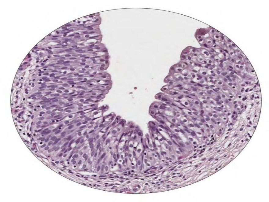
The type of epithelium as shown in the image below is present in .
A) Duodenum
B) Common bile duct
C) Skin
D) Urinary bladder
Correct Answer:D
Explanation:
The type of epithelium shown in the image is the transitional epithelium and it is present in the urinary bladder.
Transitional epithelium is a specialized epithelium lining most of the urinary tract, hence it is also called the urothelium. The deepest layer consists of columnar or cuboidal cells, and the middle layer consists of polyhedral or pear-shaped cells. The surface layer consists of large, polypoid, dome-shaped, or umbrella-shaped cells. As the epithelium gets stretched to accommodate urine, the surface cells become flattened. The cells are called transitional due to this apparent change (transition) from stratified cuboidal epithelium in a relaxed state to stratified squamous epithelium when it is stretched.
The cavity in the shaft of the fetal long bone mainly contains .
Image not available for this question yet.
A) Red
B) Yellow
C) Gelatinous
D) White
Correct Answer:A
Explanation:
The cavity in the shaft of the fetal long bone mainly contains red .
Bone is a soft, pulpy tissue found in the cavity and the spaces of spongy bone. It is mainly of two types:
Yellow - made up predominantly of fat cells and may contain few islands of hemopoietic tissue. It is seen in the shaft of the long bones of an adult.
Red - It is made up predominantly of hemopoietic tissue. It is seen throughout the skeleton of the fetus. It persists till about five years of age after which, the red in the
shaft of long bones is gradually replaced by the yellow . By around 20-25 years of age, the sites where the red persists include:
Vertebrae
Sternum
Ribs
Clavicles
Scapulae
Pelvis
Cranial bones
Proximal ends of femur and humerus
The term gelatinous refers to the degenerated bone in the cranial bones, seen in old age. There is no white .
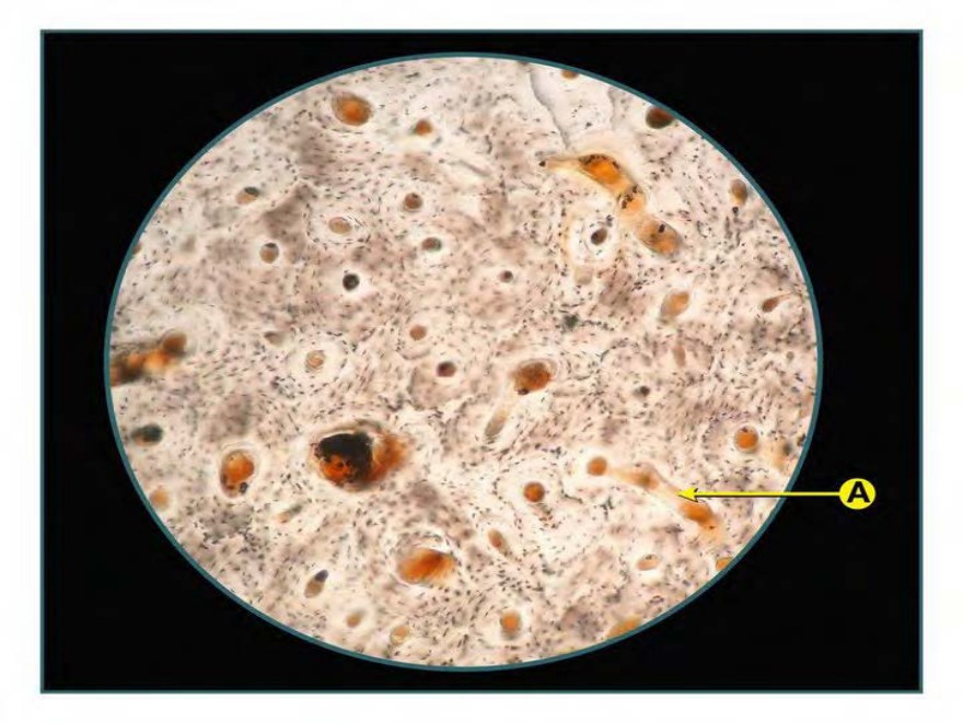
The image shows the microscopic appearance of the lamellar bone. Identify the structure marked as A.
A) Volkmann's canal
B) Haversian canal
C) Interstitial lamellae
D) Circumferential lamellae
Correct Answer:A
Explanation:
The structure marked as A in the above image is Volkmann's canal. The Haversian canals communicate with each other by the transverse perforating canals, which are called Volkmann's canals.
The Haversian system or osteon constitute most part of the compact bone. Each Haversian system contains a central canal within which the small blood vessels, nerves, and endosteum are present. This canal is surrounded by multiple layers or lamellae of the calcified matrix containing type I collagen fibers. These canals communicate with each other by Volkmann's canal.
Successive lamellae are separated from each other by lacunae containing osteocytes. These osteocytes are in communication with each other, the central canal and the periphery of the osteon via the dendritic processes present within the canaliculi. The parallel lamellae present between osteons is called interstitial lamellae and the parallel lamellae present near the periosteum are called the circumferential lamellae.
On microscopy, osteon can be identified by
Presence of Haversian canal with concentric lamellae around it
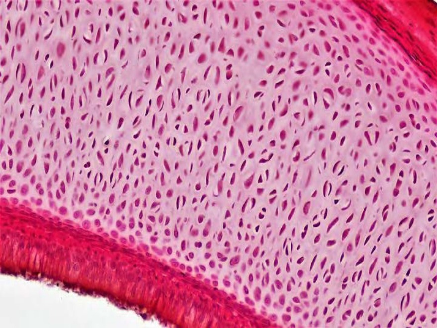
The type of cartilage as shown in the image is present in 214
A) Terminal bronchioles
B) Intervertebral discs
C) Bronchi
D) Epiglottis
Correct Answer:C
Explanation:
The type of cartilage shown in the above image is hyaline cartilage and it is present in bronchi.
Hyaline cartilage is made up of chondrocytes and matrix. The chondrocytes in the deeper matrix are rounded in shape and may appear in a group of eight that originate by the mitotic divisions of a single chondroblast. These groups of cells are called isogenous cell aggregates or cell nests. The important components of the matrix are type II collagen, aggrecan (proteoglycan) and chondronectin (glycoprotein). The matrix which immediately surrounds the chondrocytes is basophilic and is known as the territorial matrix and the matrix which are more distant from the cells appears pale and is known as the interterritorial matrix.
On microscopy, hyaline cartilage can be identified by:
Presence of chondrocytes within lacunae.
Group of two or more chondrocytes which are known as isogenous cell aggregates or cell nests.
Basophilic territorial matrix and pale interterritorial matrix
Presence of perichondrium with outer fibrous and the inner cellular layer