What is a part of the embryo that exerts morphogenetic stimulus on its adjacent part called?
Image not available for this question yet.
A) Differentiator
B) Organizer
C) Stem cell
D) Inducer
Correct Answer:B
Explanation:
Any part of the embryo that exerts a morphogenetic stimulus on adjacent parts is known as the organizer.
First level is the primary organizer that induces the development of new tissues. This newly developed tissue can then induce another tissue in turn to become the secondary organizer. Therefore, there is a sequence or chain of organizers called primary, secondary, and tertiary organizers.
Examples of the three levels of organizers:
Primary organizer: e.g., Blastopore induces differentiation of notochord
Secondary organizer: e.g., Notochord induces the development of the neural tube (that forms the brain and spinal cord)
Tertiary organizer. e.g., Neural tube induces segmentation of paraxial mesoderm into somites
Which of the following represents the embryonic period of development?
Image not available for this question yet.
A) 0-2 weeks
B) 3-8 weeks
C) 8-10 weeks
D) 10-12 weeks
Correct Answer:B
Explanation:
The embryonic period extends from the 3rd week of intrauterine life to the 8th week of intrauterine life.
The prenatal period is divided into 3 different stages:
Stages of prenatal developme nt
Pre-embryonic period(First 2 weeks)
Embryonic period(3-8 weeks)
Fetal period(9 weeks to birth)
Important events of each stag e
FertilizationCleavage and bla stocyst formationImplantatio nFormation of embryoblast a nd trophoblastDifferentiation of embryoblast totwo-layere d (bilaminar) germ discDiffer entiation of trophoblast into cytotrophoblast and syncytiot rophoblast
Trilaminar germ discformati on with the formation of 3 ge rm layersEarly organogenesis Formation of extraembryonic supportive organs and mem branes such as placenta, umb ilical cord, amnion, and allan tois
The spermatozoa are produced in seminiferous tubules by a process known as spermatogenesis. In this process, spermatogonia are transformed into spermatozoa.
Shortly before puberty, the sex cords acquire a lumen and become seminiferous tubules,
and primordial germ cells differentiate into spermatogonial stem cells. These spermatogonial stem cells then give rise to type A spermatogonia. This marks the beginning of spermatogenesis.
Which stage of spermatogenesis is characterized by an independent assortment of maternal and paternal chromosomes?
Image not available for this question yet.
A) Spermatogonia to primary spermatocyte
B) Primary spermatocyte to secondary spermatocyte
C) Secondary spermatocyte to spermatid
D) Spermatid to spermatozoa
Correct Answer:B
Explanation:
Independent assortment is the random distribution of chromosomes. It takes place during meiosis I where there is division from primary spermatocyte to secondary spermatocyte.
The law of independent assortment states that alleles for separate traits are passed independently of one another into the gametes.
During the first meiotic division, paternal and maternal chromosomes are distributed between the daughter cells entirely at random. This prevents all chromosomes from the same source (mother or father) from ending up in the same gamete. After meiosis, each haploid daughter cell contains a mixture of genes from the mother and father.
Spermatozoa attain full motility during their passage through the epididymis.
Initially, spermatozoa are only slightly motile, but they obtain full progressive motility in the epididymis. This involves the activation of a unique set of cation channel proteins from the CatSper family located in the principal piece of the sperm tail.
A 30-year-old woman came to the obstetrician for in-vitro fertilization. FSH analogs were injected to induce follicular growth and hCG was injected to induce final oocyte maturation. The oocyte was then retrieved transvaginally. The oocyte thus retrieved is arrested at which of the following stages?
A) Prophase of meiosis
B) Prophase of meiosis II
C) Metaphase of meiosis
D) Metaphase of meiosis II
Correct Answer:D
Explanation:
The oocyte retrieved after final maturation is the secondary oocyte that is arrested in metaphase of meiosis II.
Steps of oogenesis after puberty are shown in the image below:
If there is no fertilization, the secondary oocyte fails to complete the second meiotic division and degenerates about 24 hours after ovulation.
In in-vitro fertilization, initially, FSH analogs are administered to induce follicular growth. At this stage, the oocyte is arrested in the prophase stage of meiosis I. The final maturation of the oocyte is induced by the administration of hCG. This causes the oocyte to resume the meiotic division, complete meiosis I, and enter meiosis II. However, the oocyte is arrested in the metaphase of meiosis II until fertilization.
Q607.
Anatomy
Medium
4m
Image missing
Topic: Pre-Embryonic Phase of DevelopmentSource: Internal
Explanation ready
Which of the following is included in pre-embryonic phase of development?
Image not available for this question yet.
A) First 2 weeks of intrauterine development
B) First 3 weeks of intrauterine development
C) First 4 weeks of intrauterine development
D) First 8 weeks of intrauterine development
Correct Answer:A
Explanation:
The pre-embryonic phase includes the first 2 weeks of intrauterine development, from fertilization to the formation of bilaminar germ disc.
Major morphogenetic events that occur in the pre-embryonic phase include:
Fertilization
Cleavage
Transportation of cleaving zygote
Formation of blastocyst
Implantation
Specialization of primordial embryonic tissue
Differentiation of embryoblast to bilaminar germ disc
Differentiation of trophoblast to cytotrophoblast and syncytiotrophoblast
Q608.
Anatomy
Medium
4m
Image missing
Topic: Pre-Embryonic Phase of DevelopmentSource: Internal
Explanation ready
What is the final step of maturation of spermatozoon before fertilization known as?
Image not available for this question yet.
A) Spermiogenesis
B) Capacitation
C) Acrosome reaction
D) Cortical reaction
Correct Answer:B
Explanation:
The final step of maturation of spermatozoon before fertilization is known as capacitation.
Capacitation involves a period of conditioning of the sperm in the female reproductive tract, predominantly in the uterine tube. It provides the sperms with the ability to penetrate the outer layer of the ovum. Chemical changes in the tail provide them with greater mobility. It lasts approximately 7 hours.
Q609.
Anatomy
Medium
4m
Image missing
Topic: Pre-Embryonic Phase of DevelopmentSource: Internal
Explanation ready
A 26-year-old man presented with sudden onset of visual loss. After evaluation, he was diagnosed with a mitochondrial disorder called Leber's hereditary optic neuropathy. What is the origin of the affected DNA in this condition?
Image not available for this question yet.
A) Paternal only
B) Maternal only
C) 50 paternal and 50 maternal
D) Either paternal or maternal
Correct Answer:B
Explanation:
Mitochondrial disorders (e.g., Leber's hereditary optic neuropathy) have mutations in the mitochondrial DNA. Mitochondria and mitochondrial DNA of all human adult cells are of maternal origin.
During fertilization, the sperm's midpiece (with mitochondria) and tail are discarded. The sperm nucleus becomes the male pronucleus. Therefore, only the mitochondria present within the secondary oocyte (maternal mitochondria) remain in the fertilized zygote. Mitochondria, mitochondrial genetic variations, and mutations are passed only through the female line.
Q610.
Anatomy
Medium
4m
Image missing
Topic: Pre-Embryonic Phase of DevelopmentSource: Internal
Explanation ready
What does the inner cell mass form during blastocyst formation?
Image not available for this question yet.
A) Cytotrophoblast
B) Syncytiotrophoblast
C) Embryoblast
D) Blastocele
Correct Answer:C
Explanation:
The inner cell mass gives rise to the embryoblast, which forms the embryo proper.
Around the 4th-5th day of development, the morula becomes the blastocyst. Fluid passes into the morula from the uterine cavity and converts it to a blastocyst with a fluid-filled cavity called blastocoele. This fluid partially separates the cells of the inner cell mass from the outer cell mass.
Cell masses in the blastocyst:
The outer cell mass is called the trophoblast and forms a major part of the placenta
The inner cell mass is called the embryoblast and forms the embryo proper
Q611.
Anatomy
Medium
4m
Image missing
Topic: Pre-Embryonic Phase of DevelopmentSource: Internal
Explanation ready
Arrange the following events of pre-embryonic development in order, from the earlier stage to the later stage.
Image not available for this question yet.
A) 2 – 4 – 1 – 3
B) 3 – 1 – 2 – 4
C) 4 – 2 – 3 – 1
D) 4 – 2 – 1 – 3
Correct Answer:D
Explanation:
The correct order of the listed events of the pre-embryonic period is cleavage - compaction -
cavitation - implantation.
Events of the pre-embryonic phase of development:
Fertilization
Cleavage - Formation of blastomeres; the cells are loosely arranged until the 8-cell stage.
Compaction - After 3 cleavages, the blastomeres are held together by tight junctions.
Differentiation - The compacted 8-cell stage divides to form a 16-cell morula.
Cavitation - Formation of the blastocyst with inner cell and outer cell mass.
Hatching of the blastocyst - Release of the blastocyst from the zona pellucida.
Implantation - Attachment of developing embryo to the uterine endometrium.
Cell mass differentiation - Differentiation of the embryoblast (inner cell mass) into columnar epiblast cells and cuboidal hypoblast cells.
Bilaminar disc formation - Formation of the bilaminar disc suspended by the connecting stalk. It has two layers (epiblast and hypoblast) and two cavities (amniotic and yolk sac).
Q612.
Anatomy
Medium
4m
Image missing
Topic: Embryonic Phase of DevelopmentSource: Internal
Explanation ready
Which of the following germ layer(s) are formed from the epiblast?
Image not available for this question yet.
A) Ectoderm
B) Endoderm, Mesoderm, and Ectoderm
C) Ectoderm and Mesoderm
D) Endoderm and Mesoderm
Correct Answer:B
Explanation:
Epiblast gives rise to all the 3 germ layers through the process of gastrulation.
After the formation of the primitive streak, the epiblast cells migrate toward the primitive streak. When they arrive at the primitive streak, these cells become flask-shaped, detach from the epiblast, and slip beneath the primitive streak. This inward movement is known as invagination.
Some of the invaginated epiblast cells displace the hypoblast to create the embryonic endoderm. Endoderm is the first germ layer to form.
Some of the invaginated cells then start occupying the space between the epiblast and newly created endoderm to form mesoderm. Cells remaining in the epiblast then form the ectoderm.
The image given below shows the formation of germ layers.
Q613.
Anatomy
Medium
4m
Image missing
Topic: Embryonic Phase of DevelopmentSource: Internal
Explanation ready
Arrange the following structures formed during the development of the definitive notochord, from the earlier stage to the later stage.
Image not available for this question yet.
A) 3 – 4 – 2 – 1
B) 1 – 4 – 2 – 3
C) 3 – 2 – 4 – 1
D) 1 – 2 – 4 – 3
Correct Answer:C
Explanation:
The correct order is blastopore - notochordal process - notochordal canal - notochordal plate -
definitive notochord. Notochord formation steps:
• Primitive knot - The cranial end of the primitive streak thickens to form the primitive knot
(primitive node or Hensen's node).
Primitive pit (Blastopore) - A depression appears in the center of the knot known as the primitive pit (or blastopore).
Notochordal process - The cells in the primitive knot multiply and pass cranially in the midline between the ectoderm and endoderm. They reach up to the caudal margin of the prechordal plate.
These cells form a solid chord known as the notochordal process or head process.
Notochordal canal - The cavity of the blastopore then extends into the notochordal process to form a tube-like structure called the notochordal canal.
Notochordal plate - The floor of the notochordal canal gets intercalated (mixed up) with the endoderm and gradually breaks down. This eventually leads to the formation of a flat plate-like structure notochordal plate.
Definitive notochord - The cells from the notochordal plate proliferate to form a solid rod-like structure called a definitive notochord.
Q614.
Anatomy
Medium
4m
Image missing
Topic: Embryonic Phase of DevelopmentSource: Internal
Explanation ready
Which of the following statements regarding the notochord is false?
Image not available for this question yet.
A) It induces neural plate formation
B) It persists as apical ligament of the dens
C) It persists as nucleus pulposus
D) It develops from hypoblast cells
Correct Answer:D
Explanation:
The notochord is derived from the epiblast and not from the hypoblast cells.
Epiblast cells from the primitive knot region of the primitive streak multiply and pass cranially in the midline between the ectoderm and endoderm. They undergo several stages of rearrangement to form the notochord.
During the initial stages of neurulation, the notochord plays a role in the induction of overlying ectoderm to thicken and form the neural plate.
Although the notochord regresses, it has 2 important remnants:
Nucleus pulposus in the region of the intervertebral disc
Apical ligament of the dens of axis vertebra
Q615.
Anatomy
Medium
4m
Image missing
Topic: Embryonic Phase of DevelopmentSource: Internal
Explanation ready
A newborn presented to the NICU with respiratory distress and meconium discharge from the umbilicus. The condition occurs due to the patency of the duct that connects the primitive gut to the .
Image not available for this question yet.
A) Embryonic body cavity
B) Chorionic cavity
C) Amniotic cavity
D) Yolk sac
Correct Answer:D
Explanation:
The presence of meconium discharge at the umbilicus is suggestive of vitelline fistula. It occurs due to patent vitelline duct, which connects the primitive gut to the yolk sac.
The primitive gut is connected to the definitive yolk sac through a narrow stalk-like communication known as the vitellointestinal duct. It is also known as vitelline duct or yolk stalk or omphalomesenteric duct.
Due to the folding of the embryo in the cephalocaudal and lateral directions, the embryo
gets enclosed all around by ectoderm, except in the region through which the vitellointestinal duct passes. In this region, there is a circular aperture, which is now called the umbilical opening. The vitellointestinal duct completely obliterates by the 7th week of development.
Vitelline duct abnormalities:
Meckel's diverticulum: In about 2 o the individuals, a portion of the vitelline duct persists an outpouching of the ileum called, Meckel diverticulum. The diverticulum is usually asymptomatic unless it contains heterotopic pancreatic or gastric mucosa, which may lead to bleeding, ulceration, or perforation.
Vitelline cyst: In some individuals, both the ends of the vitelline duct transforms into fibrous cords. However, the middle portion of the duct forms a large cyst called, enterocystoma, or vitelline cyst. As the fibrous cords run across the peritoneal cavity, it may lead to intestinal obstruction, strangulation, or volvulus.
Q616.
Anatomy
Medium
4m
Image ready
Topic: Placenta, Fetal Membranes and TwinningSource: Internal
Explanation ready
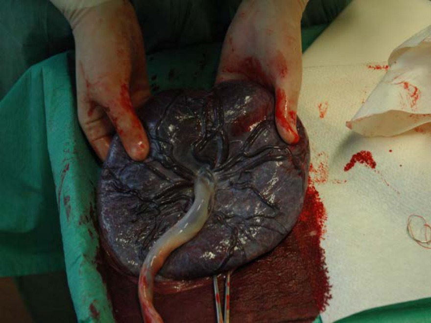
Which of the following contributes to the maternal and fetal components of the structure shown below, respectively? 46
A) Decidua basalis and chorion laeve
B) Decidua capsularis and chorion frondosum
C) Decidua capsularis and chorion laeve
D) Decidua basalis and chorion frondosum
Correct Answer:D
Explanation:
The image shown is of a placenta. The maternal component of the placenta is from the decidua basalis and the fetal component is from the chorion frondosum.
The placenta is a fetomaternal organ containing tissues from two different sources as follows:
Maternal component - contributed by the decidua basalis. The decidua basalis is also referred to as the decidua serotina or decidual plate.
Fetal component - contributed by chorion frondosum. The chorionic villi adjacent to the decidua basalis undergo significant development and branching to form a bushy region called chorion frondosum.
Q617.
Anatomy
Medium
4m
Image missing
Topic: Placenta, Fetal Membranes and TwinningSource: Internal
Explanation ready
Which of the following are components of the definitive chorion?
Image not available for this question yet.
A) Cytotrophoblast and syncytiotrophoblast
B) Extraembryonic somatic mesoderm and cytotrophoblast
C) Extraembryonic somatic mesoderm and syncytiotrophoblast
D) Extraembryonic somatic mesoderm, cytotrophoblast, and syncytiotrophoblast
Correct Answer:D
Explanation:
The definitive chorion is an extraembryonic membrane composed of three components:
Extraembryonic somatic mesoderm
Cytotrophoblast
Syncytiotrophoblast
It contributes to the fetal portion of the placenta, including the villi and villus lakes.
Q618.
Anatomy
Medium
4m
Image ready
Topic: Placenta, Fetal Membranes and TwinningSource: Internal
Explanation ready
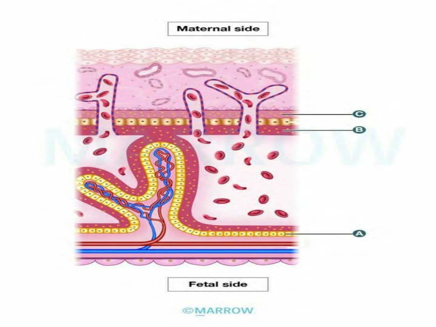
Identify the layers marked as A, B, and C, respectively, in the image of the definitive placenta.
A) Rohr's layer, Langhan's layer, Nitabuch's layer
B) Langhan's layer, Rohr's layer, Nitabuch's layer
C) Langhan's layer, Nitabuch's layer, Rohr's layer
The structures marked in the above image are (A) Langhan's layer, (B) Rohr's layer, and (C)
Nitabuch's layer.
The definitive placenta consists of the chorionic plate on the fetal aspect and a basal plate on the maternal aspect with the intervillous space between them.
The structures of the definitive placenta are shown in the image below:
Q619.
Anatomy
Medium
4m
Image missing
Topic: Placenta, Fetal Membranes and TwinningSource: Internal
Explanation ready
Identify the structures marked as 1 to 4, respectively, in the chorionic plate of the placental tissue as depicted below. 48
Image not available for this question yet.
A) Amnion, extraembryonic mesoderm, syncytiotrophoblast, cytotrophoblast
B) Extraembryonic mesoderm, amnion, syncytiotrophoblast, cytotrophoblast
C) Amnion, extraembryonic mesoderm, cytotrophoblast, syncytiotrophoblast
The structures labeled in the above image of the chorionic plate (fetal component) are (1) amnion,
(2) extraembryonic mesoderm, (3) cytotrophoblast, and (4) syncytiotrophoblast.
Q620.
Anatomy
Medium
4m
Image missing
Topic: Placenta, Fetal Membranes and TwinningSource: Internal
Explanation ready
Which of the following is present in the intervillous space of placenta?
Image not available for this question yet.
A) Maternal blood
B) Fetal blood
C) Maternal and fetal blood
D) Amniotic fluid
Correct Answer:A
Explanation:
The intervillous space between the chorionic plate and the basal plate of the placenta contains only maternal blood. Fetal blood circulates in the blood vessels in the villi.
Maternal blood flows into the lacunar spaces of the syncytiotrophoblast as early as the 9th day of pregnancy. After the formation of chorionic villi, the lacunar space is known as the intervillous space.
Eroded uterine spiral arteries empty into the intervillous space and immerse the chorionic villi in maternal blood. In the fully formed placenta, the intervillous spaces contain about 150 mL of blood that is replaced every 15–20 seconds (i.e. three to four times per minute).
Q621.
Anatomy
Medium
4m
Image ready
Topic: Placenta, Fetal Membranes and TwinningSource: Internal
Explanation ready
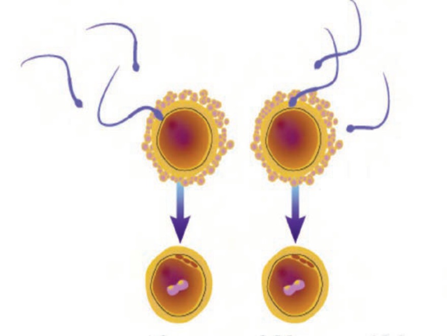
Fertili‹ation, as shown in the image, will result in twins that are
A) Di‹ygotic, Bichorial, Biamniotic
B) Di‹ygotic, Monochorial, Biamniotic
C) Mono‹ygotic, Monochorial, Biamniotic
D) Mono‹ygotic, Monochorial, Monoamniotic
Correct Answer:A
Explanation:
The above image shows the fertilization of two different ova by two different sperms that will result in dizygotic twins, each with its own chorion and amnion (bichorial, biamnionic).
Dizygotic twins arise from two oocytes that mature during the same menstrual cycle and are fertilized by two different sperm cells. Thus, dizygotic twins can be of the same or opposite sex.
Dizygotic embryos implant separately and develop membranes that are independent of each other. Each twin has its own placenta, its own chorion, and its own amniotic cavity.
Dizygotic twins are more common than monozygotic twins. They are also known as fraternal twins, sibling twins, or false twins because genetically they are as different as two separately born siblings of the same biological parents.
The image given below shows how fertilization occurs in fraternal and identical twins.
Q622.
Anatomy
Medium
4m
Image ready
Topic: Placenta, Fetal Membranes and TwinningSource: Internal
Explanation ready
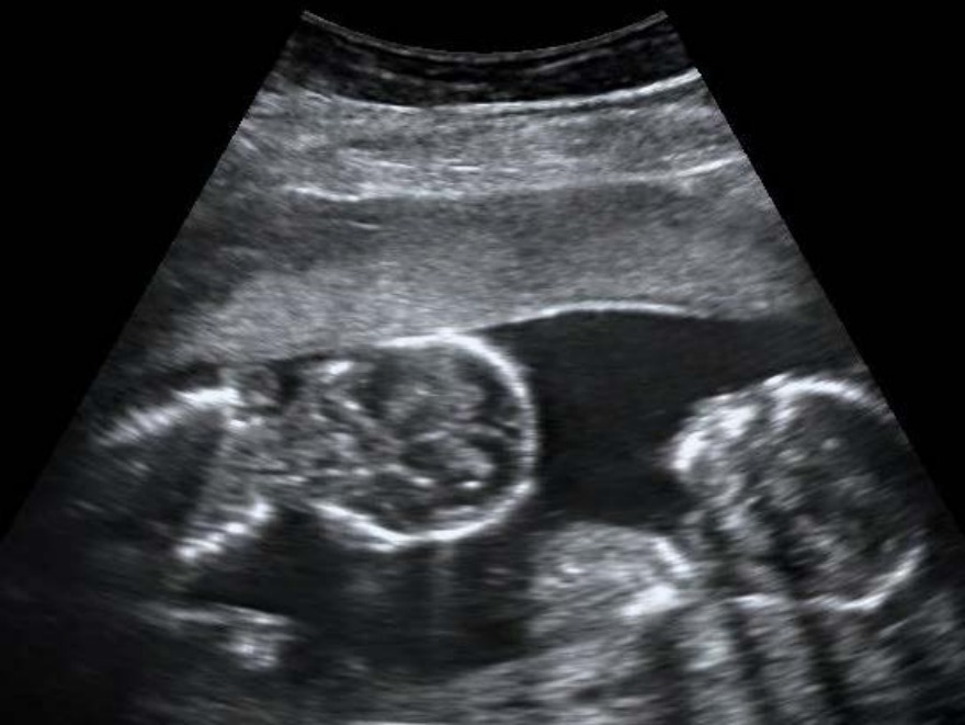
Œn USç examination of a pregnant woman at 1Ž weeks gestation, the following finding was noted. Which of the following events will result in the development of this condition? 50
A) Early blastomere separation
B) Duplication of inner cell mass before amniotic sac formation
C) Duplication of embryonic disc between 4 -è days after fertili‹ation
D) Duplication of embryonic disc between ê - 1‘ days after fertili‹ation
Correct Answer:D
Explanation:
The USG examination is suggestive of monozygotic, monochorionic, monoamniotic
twins. Duplication of the embryonic disc between the 8-12 days after fertilization will result in the development of this condition.
At this stage, the amniotic sac is already formed. In this case, the two fetuses share a common chorion and common amniotic cavity.
The image given below shows a diagrammatic representation of the placentation.
Q623.
Anatomy
Medium
4m
Image ready
Topic: Placenta, Fetal Membranes and TwinningSource: Internal
Explanation ready
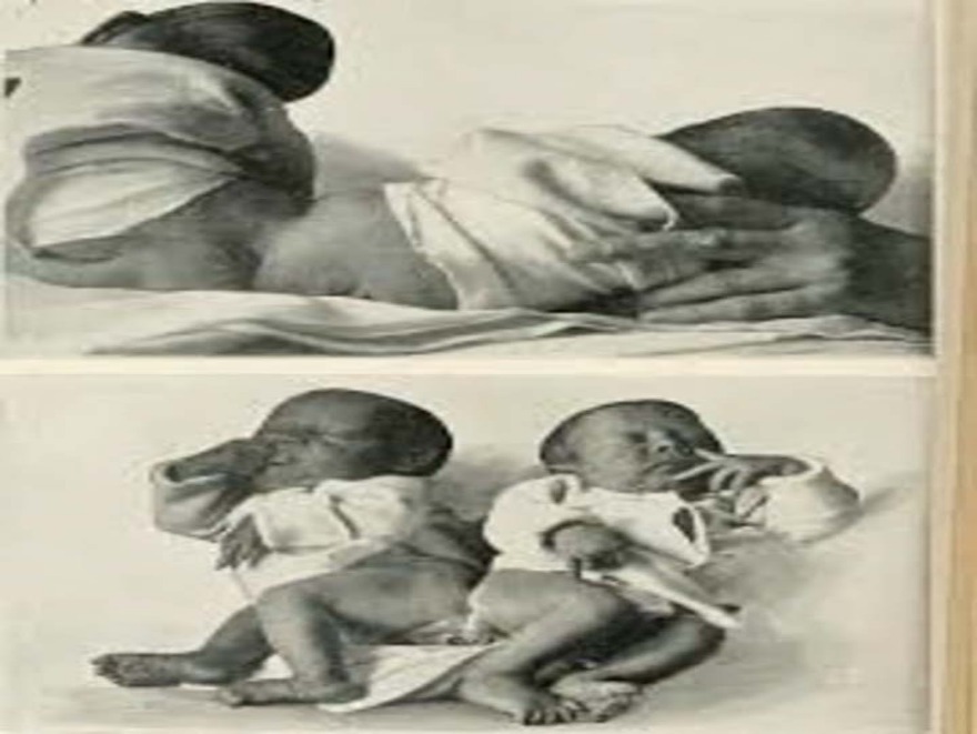
The following finding was noted in the twins that were delivered a couple of hours ago. Identify the condition.
A) Heteropagous
B) Xiphipagous
C) Omphalopagous
D) Pygopagus
Correct Answer:D
Explanation:
The image shows conjoined twins with fusion in the sacral region, which is known as the pygopagus.
In monochorionic monoamniotic twinning, incomplete duplication of the embryonic disc can occur. Incomplete separation of the monozygotic twins results in the birth of two infants that are joined or fused together at some part of the body. These twins are known as conjoined twins or Siamese twins.
Depending on the degree of incomplete separation, different types of conjoined twins can form:
Q624.
Anatomy
Medium
4m
Image missing
Topic: Placenta, Fetal Membranes and TwinningSource: Internal
Explanation ready
A 37-year-old man is diagnosed with renal failure and is advised to undergo renal transplantation. The patient has a twin brother who is willing to donate his kidney. Which of the following types of twins are HLA identical, hence the recepient twin does not require immunosuppression?
Image not available for this question yet.
A) 1 only
B) 2 and 3
C) 3 only
D) 1, 2, and 3
Correct Answer:D
Explanation:
All monozygotic twins are of the same gender, genetically identical, have identical HLA genes, regardless of the stage of separation of the cell mass.
All monozygotic twins develop from a single zygote that is formed as a result of the fertilization of a single oocyte with a single sperm. At a later stage, there is a separation of the blastomeres, inner cell mass, or embryonic disc to give rise to twins. Since they are formed from a single zygote, the twins have similar genetic composition.
A 4-day-old infant who was delivered at home is brought to the pediatrician for a routine checkup. The infant has a noticeable small mandible and cleft palate. A CT scan reveals hypoplasia of the mandible and a defect in the middle ear cavity and tympanic membrane. A developmental defect of which of the following pharyngeal arches is most likely? 63
Image not available for this question yet.
A) 1st pharyngeal arch
B) 2nd pharyngeal arch
C) 3rd pharyngeal arch
D) 6th pharyngeal arch
Correct Answer:A
Explanation:
The clinical features are suggestive of first arch syndrome due to the defect in the development of the first pharyngeal arch.
The derivatives of the first pharyngeal arch are:
Skeletal derivatives: It has 2 prominences -
Mandibular prominence (ventral)
Meckel's cartilage: Malleus, incus, anterior malleolar ligament, the spine of the sphenoid, and the sphenomandibular ligament.
Mesenchyme: In the jaw region, it surrounds the Meckel's cartilage and undergoes membranous ossification to form the mandible
Maxillary prominence (dorsal)
Mesenchyme: Maxilla, zygomatic bone, and part of the temporal bone are formed by membranous ossification