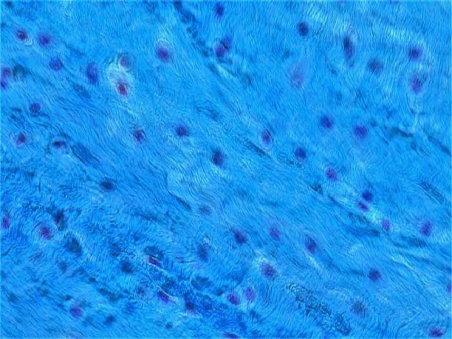
Which of the following statements is true about the type of cartilage shown in the histological image below?
A) It is present in epiphyseal plate
B) Consists of thin collagen fibres
C) Not covered with perichondrium
D) It is present in bronchi
Correct Answer:C
Explanation:
The above image shows a section of fibrocartilage stained with trichrome stain. Fibrocartilage is not covered by perichondrium.
The fibrocartilage is an intermediate between dense fibrous connective tissue (tendon and ligaments) and hyaline cartilage. It consists of bundles of thick collagen fibres (both type I and II) with rows of chondrocytes present between the bundles.
On microscopy, fibrocartilage can be identified by:
Presence of chondrocytes
Matrix containing abundant collagen fibres
Options A amp; D: Both the epiphyseal plate and bronchi have the hyaline type of cartilage.
Note: The colour of cartilage should not be considered while deciding its type, as it largely depends on the type of stain used. Instead, the structure or organisation of the cartilage tissue should be considered.
Each bundle of muscle fibres is surrounded by the perimysium. Organization of skeletal muscle:
Muscle fibre or myocyte - made up of myofibrils and consists of several elongated nuclei, which lie along the periphery of the fibre just under the cell membrane. The cell membrane of the muscle fibre is called the sarcolemma, while the cytoplasm is called sarcoplasm. Each muscle fibre is surrounded by a delicate connective tissue called endomysium.
Fascicle - Group of muscle fibres together form fascicle. Each fascicle is surrounded by the perimysium. The perimysium serves as a pathway for nerves and blood vessels.
Muscle - Group of fascicles together form a skeletal muscle. The entire skeletal muscle is surrounded by epimysium. Epimysium contains larger nerves, blood vessels and lymphatics of the muscle.
Q679.
Anatomy
Medium
4m
Image missing
Topic: Tongue and PalateSource: Internal
Explanation ready
While testing the taste sensation in a stroke patient, you place a few drops of sugar water over the circumvallate papillae. Which of the following nerves will carry the taste pathway here?
Image not available for this question yet.
A) Chorda tympani
B) Greater petrosal nerve
C) Internal laryngeal nerve
D) Glossopharyngeal nerve
Correct Answer:D
Explanation:
The taste pathway for circumvallate papillae of the tongue goes through the glossopharyngeal nerve.
Taste sensation from the anterior 2/3rd of the tongue except the circumvallate papillae is carried by the chorda tympani branch of the facial nerve.
Areas innervated by glossopharyngeal nerve:
The circumvallate papillae
Posterior 1/3rd of the tongue
Palatoglossal arches
The oropharynx
Taste buds in the extreme posterior part of the tongue and epiglottis receive fibres from the internal laryngeal branch of the vagus.
Q680.
Anatomy
Medium
4m
Image missing
Topic: Tongue and PalateSource: Internal
Explanation ready
A patient presents with a painful aphthous ulcer at the tip of the tongue. Which of the following nerves gives rise to the branch carrying this sensation?
Image not available for this question yet.
A) Glossopharyngeal nerve
B) Chorda tympani nerve
C) Mandibular nerve
D) Vagus nerve
Correct Answer:C
Explanation:
Pain sensation at the tip of the tongue is carried by the lingual nerve which is a branch of the mandibular nerve.
Q681.
Anatomy
Medium
4m
Image missing
Topic: Tongue and PalateSource: Internal
Explanation ready
A 50-year-old man with a history of tobacco chewing presented with a painless ulcer over the tip of the tongue. Wedge biopsy revealed squamous cell carcinoma. Which of the following lymph nodes would you first biopsy?
Image not available for this question yet.
A) Submandibular lymph nodes
B) Submental lymph nodes
C) Deep cervical lymph nodes
D) Jugulo-omohyoid lymph nodes
Correct Answer:B
Explanation:
The submental lymph node will be biopsied first as it is the sentinel lymph node. The sentinel node is the first node that the tumor site drains into.
In squamous cell carcinoma of the tongue, submental lymph nodes are involved in lesions of the tip and submandibular lymph nodes are involved in lesions of the sides of the tongue. Bilateral neck lymph node spread is common due to the crossing of lymph vessels in the tongue.
Lymphatic drainage of the to ngue
Marginal
Lateralmargin of the tongue Central
Dorsal
Area
Tipof the tongue
Unilateralsubmandibularnod es
Central part of the anterior 2/ 3rd of the tongue
Posterior 1/3rd of the tongue and circumvallate papillae
Lymph nodes
Bilateral submental nodes
Bilateraljugulodigastric and j ugulo-omohyoidnodes
Bilateraljugulodigastricandju gulo-omohyoidnodes
Q682.
Anatomy
Medium
4m
Image missing
Topic: Tongue and PalateSource: Internal
Explanation ready
The sensory fibers from the taste buds in the hard and soft palate travel along the .
Image not available for this question yet.
A) Facial Nerve
B) Trigeminal nerve
C) Glossopharyngeal nerve
D) Vagus nerve
Correct Answer:B
Explanation:
The sensory fibers from the taste buds in the hard and soft palate travel along the trigeminal nerve.
The taste sensation from taste buds in the oral surface of the soft palate is carried by the lesser palatine nerve.
The taste fibers initially travel in the greater petrosal nerve (a branch of the facial nerve) and pass through the pterygopalatine ganglion without synapsing.
Q683.
Anatomy
Medium
4m
Image missing
Topic: Tongue and PalateSource: Internal
Explanation ready
Which of the following does not supply the palate?
Image not available for this question yet.
A) Tonsillar branch of facial artery
B) Ascending palatine artery
C) Descending palatine artery
D) Ascending pharyngeal artery
Correct Answer:A
Explanation:
The tonsillar branch of facial artery supplies the palatine tonsil and not the palate.
Blood supply of the palate:
Hard palate - greater (descending) palatine artery which is a branch of the third part of the maxillary artery.
Soft palate - ascending palatine branch of the facial artery, ascending pharyngeal artery, and lesser palatine arteries.
Q684.
Anatomy
Medium
4m
Image missing
Topic: PharynxSource: Internal
Explanation ready
You are an ENT resident performing endos€opy of the nasopharynx in a patient with suspe€ted nasopharyngeal €ar€inoma. Åhi€h of the following stru€tures would you not expe€t to find here?
Image not available for this question yet.
A) ‚iriform fossa
B) ‚assavant's ridge
C) ƒpening of eusta€hian tube
D) Torus tubarius
Correct Answer:A
Explanation:
Piriform fossa is a structure present in the laryngopharynx and not the nasopharynx.
The nasopharynx is present above the soft palate and is made up of the roof, floor, posterior wall, and two lateral walls. Important structures seen in the nasopharynx are:
Pharyngeal opening of the eustachian tube
Torus tubarius- tubal elevation which is present behind the opening of the eustachian tube.
çossa of Žosenmuller- A lateral pharyngeal recess that is present behind the torus tubarius. It is the most common site for nasopharyngeal carcinoma
Pharyngeal tonsil which is present in the roof and posterior wall
Passavant's ridge.
Q685.
Anatomy
Medium
4m
Image missing
Topic: PharynxSource: Internal
Explanation ready
Åhi€h of the following stru€tures forms the ‚assavant's ridge?
Image not available for this question yet.
A) „evator veli palatini and superior €onstri€tor
B) ‚alatopharyngeus and superior €onstri€tor
C) ‚alatoglossus and middle €onstri€tor
D) ‚alatoglossus and superior €onstri€tor
Correct Answer:B
Explanation:
Passavant's ridge is formed by palatopharyngeus and superior constrictor muscles.
Some fibers of the palatopharyngeus muscle pass deep to the mucous membrane of the pharynx and form an internal sphincter to the superior constrictor. These muscles contract and cause a ridge on the posterior wall of the nasopharynx which is †nown as Passavant's ridge.
Q686.
Anatomy
Medium
4m
Image missing
Topic: PharynxSource: Internal
Explanation ready
The following image shows the oropharynx in a 16-year-old girl who €ame with €omplaints of a sore throat and fever. Åhi€h of the following stru€tures will you not see in this region?
A) ‚alatopharyngeal folds
B) ‚alatine tonsils
C) ‚assavant's ridge
D) ‚alatoglossal folds
Correct Answer:C
Explanation:
The given image shows purulent inflammation of the tonsils and the case scenario is suggestive of acute tonsillitis. Passavant's ridge is not seen in the oropharynx as it is present on the posterior wall of the nasopharynx.
The oropharynx extends from below the soft palate to the upper border of the epiglottis. Structures seen in the oropharynx:
Palatoglossal folds
Palatopharyngeal folds
Palatine tonsils - between the palatoglossal and palatopharyngeal folds
Lingual tonsils
Valleculae - mucosal pouches between the tongue and the epiglottis.
Q687.
Anatomy
Medium
4m
Image missing
Topic: PharynxSource: Internal
Explanation ready
Åhere is the sinus of Šorgagni present?
Image not available for this question yet.
A) ‡etween middle €onstri€tor and inferior €onstri€tor
B) ‡etween superior €onstri€tor and middle €onstri€tor
C) ‡etween s†ull and superior €onstri€tor
D) ‡elow inferior €onstri€tor
Correct Answer:C
Explanation:
Sinus of Morgagni is a gap present between the base of the s†ull and the upper border of superior constrictor muscle.
Structures passing through the sinus of Morgagni are as follows:
Palatine branch of ascending pharyngeal artery
Ascending palatine artery
Levator veli palatini muscle
Eustachian tube
Note: Sinus of Morgagni syndrome/Trotter's triad is seen in nasopharyngeal carcinoma which spreads laterally to involve the sinus of Morgagni. It is characteriˆed by:
Conductive hearing loss
Ipsilateral immobility of the palate
Neuralgic pain.
Q688.
Anatomy
Medium
4m
Image missing
Topic: PharynxSource: Internal
Explanation ready
Åhi€h of the following mus€les is not supplied by the pharyngeal plexus? 595
Image not available for this question yet.
A) Palatoglossus
B) Stylopharyngeus
C) Middle constrictor
D) Levator veli palatini
Correct Answer:B
Explanation:
Stylopharyngeus is not supplied by the pharyngeal plexus. It is supplied by the glossopharyngeal nerve.
The pharyngeal plexus is formed by the pharyngeal branches of the glossopharyngeal nerve and the vagus nerve with contributions from the superior cervical sympathetic ganglion. It supplies:
All muscles of the soft palate
Except tensor veli palatini - supplied by the mandibular nerve
All the muscles of the pharynx
Except stylopharyngeus - supplied by the glossopharyngeal nerve.
Q689.
Anatomy
Medium
4m
Image missing
Topic: LarynxSource: Internal
Explanation ready
Which of the following laryngeal cartilages is not paired?
Image not available for this question yet.
A) Arytenoid
B) Corniculate
C) Cricoid
D) Cuneiform
Correct Answer:C
Explanation:
Cricoid is not a paired cartilage.
Q690.
Anatomy
Medium
4m
Image missing
Topic: LarynxSource: Internal
Explanation ready
Which of the following cartilages form a complete ring? 601
Image not available for this question yet.
A) Thyroid cartilage
B) Cricoid cartilage
C) Cuneiform cartilage
D) Epiglottis
Correct Answer:B
Explanation:
Cricoid cartilage forms a complete ring around the airway and this is the only laryngeal cartilage to do so.
Clinical significance:
Sellick's maneuver - Cricoid pressure is applied to occlude the esophagus without occluding the airway. It prevents regurgitation of gastric contents during intubation and rapid induction of anesthesia.
The cricoid cartilage is the narrowest part of the pediatric airway.
Q691.
Anatomy
Medium
4m
Image missing
Topic: LarynxSource: Internal
Explanation ready
Which of the following will you not list as an example of this type of cartilage?
A) Cricoid
B) Thyroid
C) Base of arytenoid
D) Apex of arytenoid
Correct Answer:D
Explanation:
The given image shows the histology of hyaline cartilage. Apex of arytenoid is made up of elastic cartilage, not hyaline cartilage.
Q692.
Anatomy
Medium
4m
Image missing
Topic: LarynxSource: Internal
Explanation ready
All of the following ligaments are components of the external laryngeal membrane except .
Image not available for this question yet.
A) Cricothyroid
B) Thyrohyoid
C) Cricotracheal
D) Hyoepiglottic
Correct Answer:A
Explanation:
Cricothyroid is the component of the intrinsic laryngeal membrane of the larynx.
The cricothyroid ligament is present between the cricoid and thyroid cartilage of the larynx. It consists of three parts: the right and the left lateral parts and the thickened median portion. The lateral parts are called conus elasticus and the median thickened part is called the median or the anterior cricothyroid ligament.
The extrinsic ligaments and membranes of the larynx are:
Thyrohyoid membrane
Hyoepiglottic ligament
Cricotracheal ligament
The intrinsic ligaments and membranes of the larynx are:
Quadrangular membrane - present between the epiglottis and the arytenoid cartilage
Cricothyroid membrane
Thyroepiglottic ligament.
Q693.
Anatomy
Medium
4m
Image missing
Topic: LarynxSource: Internal
Explanation ready
Which of the following statement is true regarding the laryngeal cavity?
Image not available for this question yet.
A) Laryngeal ventricle is between laryngeal inlet and vestibular folds
B) Vocal cords are lined by pseudostratified ciliated columnar epithelium
C) Rima glottidis is present between the vocal folds
D) Ventricle is a pouch from the anterior end of the saccule
Correct Answer:C
Explanation:
Rima glottidis is present between the vocal folds of the larynx.
The cavity of the larynx can be divided into upper, middle, and lower parts. Upper part:
It extends from the laryngeal inlet to the vestibular folds.
The laryngeal inlet is made of the upper edge of the epiglottis, aryepiglottic fold, arytenoids,
and posterior commissure.
The vestibule is the region between the laryngeal inlet and the vestibular folds.
Middle part:
It extends from rima vestibuli to the rima glottidis.
Rima vestibuli is the aperture between the two vestibular folds.
Rima glottidis is the aperture between the two vocal folds.
Ventricle is the space bounded by rima vestibuli above and rima glottidis below.
Saccule is a pouch from the anterior end of the ventricle. It contains mucous glands.
Lower part:
The lower part is also called the infraglottic cavity or subglottic cavity and extends from the
vocal cords to the lower border of the cricoid cartilage.
The larynx is covered by the pseudostratified ciliated columnar epithelium except at three sites where it is covered by non-keratinized stratified squamous epithelium. These are:
Vocal folds
Lingual surface of epiglottis
Upper part of aryepiglottic folds
Q694.
Anatomy
Medium
4m
Image missing
Topic: LarynxSource: Internal
Explanation ready
Which of the following statements is correct about the sensory innervation of the larynx?
Image not available for this question yet.
A) Above the level of vocal cords - Superior laryngeal nerve
B) Above the level of vocal cords - Recurrent laryngeal nerve
C) Below the level of vocal cords - Internal laryngeal nerve
D) Below the level of vocal cords - External laryngeal nerve
Correct Answer:A
Explanation:
The nerve supply of larynx above the level of vocal cords is by internal laryngeal nerve which is a branch of the superior laryngeal nerve.
Sensory innervation of the larynx above the level of vocal cords is by the internal laryngeal
nerve.
Sensory innervation of the larynx below the level of vocal cords is by the recurrent laryngeal nerve.
At the level of vocal cords, it is supplied both by the internal laryngeal and recurrent laryngeal nerves.
All the intrinsic muscles of the larynx are supplied by the recurrent laryngeal nerve except for cricothyroid, which is supplied by the external laryngeal nerve.
Q695.
Anatomy
Medium
4m
Image missing
Topic: LarynxSource: Internal
Explanation ready
Which among the following is false about larynx? 605
Image not available for this question yet.
A) 9 cartilages: 3 paired, 3 unpaired
B) Extends from C3-C6 vertebrae
C) External laryngeal nerve supplies all larynx muscles except cricothyroid
D) Cricothyroid is a tensor of vocal cord
Correct Answer:C
Explanation:
The external laryngeal nerve supplies the cricothyroid muscle. All other intrinsic muscles of the larynx are supplied by the recurrent laryngeal nerve.
The larynx contains three paired cartilages (arytenoid, corniculate, cuneiform) and three unpaired cartilages (epiglottis, thyroid, and cricoid).
Sometimes the triticeal cartilage (TC) may be present. This is a small, oval-shaped cartilage found within the lateral border of the thyrohyoid membrane between the greater horn of the hyoid bone and the superior horn of the thyroid cartilage (SHTC). The TC is not a constant structure; it may be present unilaterally, bilaterally, or absent.
Q696.
Anatomy
Medium
4m
Image missing
Topic: LarynxSource: Internal
Explanation ready
A surgeon is performing a thyroidectomy for a patient with a multinodular goiter. Damage to which structure is most likely to cause vocal cord paralysis?
Image not available for this question yet.
A) Recurrent laryngeal nerve
B) External laryngeal nerve
C) Internal laryngeal nerve
D) Superior laryngeal nerve
Correct Answer:A
Explanation:
Injury to the recurrent laryngeal nerve will cause vocal cord paralysis.
All the intrinsic muscles of the larynx except for the cricothyroid are supplied by the recurrent laryngeal nerve. Cricothyroid is supplied by the external laryngeal nerve which is a branch of the superior laryngeal nerve.
Since most of the laryngeal muscles are supplied by the recurrent laryngeal nerve, the most appropriate answer would be recurrent laryngeal nerve.
Q697.
Anatomy
Medium
4m
Image missing
Topic: LarynxSource: Internal
Explanation ready
A chronic smoker presented with a supraglottic growth, which was later found to be squamous cell carcinoma. How will this spread to the pre-epiglottic space?
Image not available for this question yet.
A) Lymphatic spread
B) Perforations in the epiglottis
C) Hematogeneous spread
D) Through the para-glottic space
Correct Answer:B
Explanation:
Supraglottic tumors spread to the pre-epiglottic space via the perforations in the epiglottic cartilage.
Pre-epiglottic space: It lies anterior to the epiglottis. Its relations are as follows:
Superior - Hyoepiglottic membrane median hyoepiglottic ligament
Inferior - Thyroepiglottic ligament
Anterior - Thyrohyoid membrane and median thyrohyoid ligament
Posterior - Epiglottis
Lateral - Greater cornua of the hyoid bone in the upper part, continuous with the paraglottic space in the lower part.
The laryngeal tumors can spread to the pre-epiglottic space through the multiple perforations present in the epiglottis.
Para-glottic space: This space contains the laryngeal nerve. Its relations are:
Superior - Para-glottic space is continuous with the pre-epiglottic space.
Inferior- Cricothyroid joint
Posterior- Piriform fossa
Lateral - Thyroid cartilage and thyrohyoid membrane
Medial- Quadrangular membrane in the upper part and conus elasticus in the lower part.
Q698.
Anatomy
Medium
4m
Image missing
Topic: LarynxSource: Internal
Explanation ready
606 All of the following statements are true except:
Image not available for this question yet.
A) Vagus nerve emerges via the intermediate compartment of jugular foramen
B) Galen's anastomosis refers to anastomosis between the internal laryngeal nerve and recurrent
C) Internal laryngeal nerve is accompanied by superior thyroid vessels
D) Right reccurent laryngeal nerve winds around the 1st part of subclavian artery
Correct Answer:C
Explanation:
The internal laryngeal nerve is accompanied by the superior laryngeal vessels, not the superior thyroid vessels. Along with superior laryngeal vessels, it pierces the thyrohyoid membrane.
The vagus nerve arises as several rootlets from the medulla which unite to form a single nerve and emerges via the intermediate compartment of the jugular foramen.
Galen's anastomosis is the connecting branch between the inferior laryngeal nerve (a branch of the recurrent laryngeal nerve) and the internal laryngeal nerve (a branch of the superior laryngeal nerve).
Right recurrent laryngeal nerve winds around the lower and posterior part of 1st part of the right subclavian artery and left recurrent laryngeal nerve winds around the arch of the aorta.
Q699.
Anatomy
Medium
4m
Image missing
Topic: Upper Limb Bones and JointsSource: Internal
Explanation ready
Which is the first bone to ossify in the human body?
Image not available for this question yet.
A) Humerus
B) Clavicle
C) Scapula
D) Lunate
Correct Answer:B
Explanation:
The clavicle is the first bone to ossify in the human body. It occurs by the 5th- 6th weeks of intrauterine life.
The clavicle is a band of condensed mesenchyme in the early embryonic phase.
At the terminal ends of this band, early cartilage transformation (pre-cartilage) takes place. Intramembranous centers of ossification appear and fuse in the mesenchyme between them. Hence, it ossifies partly in the membrane and partly in cartilage.
Q700.
Anatomy
Medium
4m
Image missing
Topic: Upper Limb Bones and JointsSource: Internal
Explanation ready
Which of the following is false about clavicle?
Image not available for this question yet.
A) Ossify partly in membrane and partly in cartilage
B) Transmits part of the weight of upper limb to the axial skeleton
C) It is subcutaneous throughout its length
D) Fractures typically occur at the junction of medial 1/3rd and lateral 2/3rd
Correct Answer:D
Explanation:
Fractures of the clavicle typically occur at the junction of the medial 3/5 th and lateral 2/5 th.
Middle-third or midshaft region of the clavicle is the most common site for a fracture, especially the junction of medial 3/5 th and lateral 2/5 th.
It is the junction where the 2 primary ossification centers meet. It is also where the transition from antecurve to retro curve begins and is prone to be at high risk for fractures.
The fracture causes the upward displacement of the proximal fragment (sternocleidomastoid) and downward displacement of the distal fragment (pectoralis major and gravity).