Topic: Glands of the Head and NeckSource: Internal
Explanation ready
Match the following with the structures they supply.
Image not available for this question yet.
A) A-…, †- ‡, C-ˆ, D-‰
B) A-ˆ, †-‰, C-‡, D-…
C) A-ˆ, †-‡, C-‰, D-…
D) A-…, †-‡, C-‰, D-ˆ
Correct Answer:C
Explanation:
The otic ganglion supplies the parotid gland.
The pterygopalatine ganglion (also known as Meckel's ganglion) supplies the lacrimal glands and palatal glands
The submandibular ganglion supplies the submandibular glands and sublingual glands.
Q552.
Anatomy
Medium
4m
Image missing
Topic: Glands of the Head and NeckSource: Internal
Explanation ready
557 A medical student skipped breakfast to be able to attend his morning class. He is currently thinking about having food and coffee after class, and notices increased salivary secretion. The nerve fibres to the parotid gland responsible for this come from all except:
Image not available for this question yet.
A) Otic ganglion
B) Greater petrosal nerve
C) Auriculotemporal nerve
D) Tympanic plexus
Correct Answer:B
Explanation:
The given case scenario is suggestive of parasympathetic stimulation of the salivary glands, causing the parasympathetic secretomotor fibres to increase salivary secretion.
The parasympathetic fibres to the parotid gland are carried by the lesser petrosal nerve.
Otic ganglion is a parasympathetic ganglion which supplies secretomotor fibres to the parotid gland and is present below the foramen ovale.
Preganglionic fibres originate in the inferior salivatory nucleus and pass through the lesser petrosal nerve which is the continuation of the tympanic branch of the glossopharyngeal nerve.
They relay in the otic ganglion.
Postganglionic fibres pass by the communicating branches to the auriculotemporal nerve and supply the parotid gland.
The greater petrosal nerve carries secretomotor fibres to the pterygopalatine ganglion and supplies the lacrimal, nasal, and palatine glands.
Sympathetic supply to the parotid gland is derived from the plexus around the middle meningeal artery.
Sensory supply to the parotid gland is from the auriculotemporal nerve and to the parotid capsule is from the sensory fibres of greater auricular nerve.
Q553.
Anatomy
Medium
4m
Image missing
Topic: Glands of the Head and NeckSource: Internal
Explanation ready
Lobes of the structure marked X are divided by
Image not available for this question yet.
A) Mylohyoid
B) Genioglossus
C) Stylohyoid
D) Styloglossus
Correct Answer:A
Explanation:
The structure marked X is the submandibular gland. Lobes of the submandibular gland are divided by the mylohyoid muscle.
The submandibular gland is divided into a larger superficial and smaller deep part by
the mylohyoid muscle. They are continuous with each other around the posterior border of the mylohyoid.
Topic: Glands of the Head and NeckSource: Internal
Explanation ready
What is the extent of the thyroid gland? 560
Image not available for this question yet.
A) B to
B) A to
C) A to
D) C to
Correct Answer:C
Explanation:
The extent of the thyroid gland is from vertebral levels A to D in the given image. It extends from vertebral levels C5 to T1.
Levels with respect to the tra cheal rings and vertebra
Lobes of the thyroid gland Isthmus of the thyroid gland
Thyroid cartilage to 4thtrach eal ringVertebral level-C5-T1
Opposite 2ndand 3rdtracheal rings
Q555.
Anatomy
Medium
4m
Image missing
Topic: Glands of the Head and NeckSource: Internal
Explanation ready
A 62-year-old woman underwent a thyroidectomy for a multinodular goiter. After coming out of anesthesia, the patient reports weakness of the right side of the body and is unable to speak. CT-brain showed multiple scattered air bubbles in the left hemisphere. Intraoperative injury to which of the following structures can lead to this condition?
Image not available for this question yet.
A) Superior or middle thyroid vein
B) Middle or inferior thyroid vein
C) Superior or inferior thyroid vein
D) Superior or inferior thyroid artery
Correct Answer:A
Explanation:
The given case scenario is suggestive of cerebral air embolism. Intra-operative injury to the superior or middle thyroid veins causes air to get sucked into them due to the negative venous pressure. The air embolus then travels up through the internal jugular vein into the cerebral venous circulation. Head-down position during surgery prevents air embolism.
Venous drainage of the thyroid gland:
Superior and middle thyroid veins - drain into the internal jugular vein
Inferior thyroid vein - drains into the brachiocephalic vein
Q556.
Anatomy
Medium
4m
Image missing
Topic: Glands of the Head and NeckSource: Internal
Explanation ready
A 40-year old female underwent total thyroidectomy and on the third postoperative day, the patient complains of numbness in her fingers and perioral area. Which of the following vessels would have been injured during the surgery?
Image not available for this question yet.
A) Anterior branch of superior thyroid artery
B) Posterior branch of superior thyroid artery
C) Superior branch of inferior thyroid artery
D) Inferior branch of inferior thyroid artery
Correct Answer:C
Explanation:
The above clinical scenario is suggestive of postoperative hypoparathyroidism due to injury to the superior branch of the inferior thyroid artery.
The parathyroid glands are small yellowish-brown structures lying between the posterior border of the thyroid gland and its capsule. There are
Two superior parathyroid glands
Two inferior parathyroid glands
These are supplied by the superior branch of the inferior thyroid artery. They secrete parathormone which helps in the regulation of the serum calcium, by the release of calcium from bones.
Q557.
Anatomy
Medium
4m
Image missing
Topic: Tongue and PalateSource: Internal
Explanation ready
All the following muscles of the tongue are derived from occipital myotomes except . 582
Image not available for this question yet.
A) Genioglossus
B) Palatoglossus
C) Styloglossus
D) Superior longitudinal muscle
Correct Answer:B
Explanation:
All the muscles of the tongue are derived from occipital myotomes except palatoglossus. It is derived from the fourth pharyngeal arch.
Hence all the muscles of the tongue are supplied by the hypoglossal nerve except the palatoglossus muscle which is supplied by the pharyngeal plexus.
Q558.
Anatomy
Medium
4m
Image missing
Topic: Tongue and PalateSource: Internal
Explanation ready
A patient with long-standing erythroplakia now presents with the following finding. Which of the following lymph nodes would be involved first in this patient? 584
Image not available for this question yet.
A) Submental lymph nodes
B) Deep cervical lymph nodes
C) Bilateral submandibular lymph nodes
D) Unilateral submandibular lymph nodes
Correct Answer:D
Explanation:
The given image shows carcinoma of the tongue involving the marginal part of the anterior 2/3rd of the tongue. Lymphatics from this region drain to unilateral submandibular lymph nodes.
All lymphatics from the tongue ultimately drain into deep cervical nodes along the internal jugular vein. Hence jugulo-omohyoid nodes are known as the lymph nodes of the tongue.
Q559.
Anatomy
Medium
4m
Image missing
Topic: Tongue and PalateSource: Internal
Explanation ready
You notice the following finding while administering OPV for a child. Which of the following nerves does not provide sensory supply to the structure involved?
Image not available for this question yet.
A) Facial nerve
B) Hypoglossal nerve
C) Glossopharyngeal nerve
D) Maxillary nerve
Correct Answer:B
Explanation:
The given image shows a cleft palate. The sensory supply to the palate is not provided by the hypoglossal nerve. It is a pure motor nerve.
General sensation from most of the soft palate is carried by:
Lesser palatine nerve (a branch of the maxillary nerve)
Pharyngeal branches of the glossopharyngeal nerve
Plexus around the tonsil (formed by tonsillar branches of the glossopharyngeal and lesser palatine nerves)
Taste sensation from the oral surface of the soft palate is carried by the lesser palatine nerve. The taste fibers initially travel in the greater petrosal nerve (a branch of the facial nerve) and pass through the pterygopalatine ganglion without synapsing.
All the muscles of the soft palate are supplied by the pharyngeal plexus (CN X), except for tensor veli palatini, which is supplied by the mandibular nerve.
The hard palate is supplied by the greater palatine and nasopalatine branches of the pterygopalatine ganglion.
Q560.
Anatomy
Medium
4m
Image missing
Topic: PharynxSource: Internal
Explanation ready
At what vertebral level is the lower border of the pharynx?
Image not available for this question yet.
A) C1
B) C2
C) C4
D) C6
Correct Answer:D
Explanation:
The lower border of the pharynx is at the level of the sixth or seventh cervical vertebrae.
The pharynx extends from the cranial base to the lower border of the cricoid cartilage (at the level of the sixth or seventh cervical vertebra). It is divided into the nasopharynx, oropharynx, and laryngopharynx.
Q561.
Anatomy
Medium
4m
Image missing
Topic: PharynxSource: Internal
Explanation ready
A …-year-old girl with pharyngitis €omplains of pain and blo€†ed sensation in her right ear. Åhere does the involved stru€ture open in the nasopharynx?
Image not available for this question yet.
A) ‡ehind middle turbinate
B) ‡ehind posterior end of inferior turbinate
C) Anterior to inferior turbinate
D) ‡etween middle and inferior turbinate
Correct Answer:B
Explanation:
The given case scenario is suggestive of a middle ear infection i.e., otitis media as a complication of pharyngitis. This is due to the spread of the infection via the eustachian tube. It opens approximately behind the posterior end of the inferior turbinate, in the lateral wall of
the nasopharynx.
The eustachian tube connects the nasopharynx and the tympanic cavity in the middle ear. It is usually collapsed and opens during activities such as yawning or swallowing. Its function is to maintain the pressure in the middle ear cavity and drain any secretions from the middle ear.
Q562.
Anatomy
Medium
4m
Image missing
Topic: PharynxSource: Internal
Explanation ready
You are an ENT resident performing indire€t laryngos€opy to visualiˆe the hypopharynx in a patient with suspe€ted glotti€ €ar€inoma. Åhi€h of the following stru€tures will you not see here?
Image not available for this question yet.
A) ‚iriform fossa
B) ‚ost €ri€oid region
C) ‰alle€ulae
D) ‚osterior pharyngeal wall
Correct Answer:C
Explanation:
Valleculae will not be seen here as they are a part of the oropharynx. èypopharynx/Laryngopharynx:
It extends from the superior border of the epiglottis to the inferior border of the cricoid cartilage.
It is separated from the oropharynx by the lateral glossoepiglottic folds.
The anterior wall is made by the parts of the laryngeal inlet, and posterior surfaces of the arytenoid and cricoid cartilages.
Pyriform fossae are present between the aryepiglottic folds medially and thyroid cartilage, thyrohyoid membrane laterally. They serve as channels that direct the solid and liquid food from the oral cavity into the esophagus.
Clinical significance: ‡ranches of the internal laryngeal nerve lie beneath the mucous membrane of the piriform fossa. èence this site can be used for infiltrating the local anesthetics.
Q563.
Anatomy
Medium
4m
Image missing
Topic: PharynxSource: Internal
Explanation ready
A 46-year-old male presented with dysphagia and regurgitation. ‡arium swallow showed the following pi€ture. Åea†ness of whi€h of the following mus€les predisposes to the given €ondition?
Image not available for this question yet.
A) ‹uperior €onstri€tor
B) Inferior €onstri€tor
C) Šiddle €onstri€tor
D) ‚alatopharyngeus
Correct Answer:B
Explanation:
The given clinical scenario with a pulsion diverticulum on the barium swallow is suggestive of Zenker's diverticulum. It originates through Killian's dehiscence, which is a site of weakness present in the inferior constrictor muscle.
The inferior constrictor muscle is made up of 2 parts:
Upper part - thyropharyngeus, made of oblique fibres.
Lower part - cricopharyngeus, made of transverse fibres.
Between the two muscles, there is a potential gap which is called the pharyngeal dimple or Killian's dehiscence.
Another site of weakness in the pharyngeal wall is the Laimer's triangle, which is present between the cricopharyngeus and the longitudinal muscle of the oesophagus.
Q564.
Anatomy
Medium
4m
Image ready
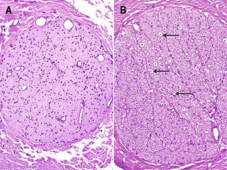
Topic: Nervous and Endocrine SystemsSource: Internal
Explanation ready
Identify the types of nerve fibers from images A and ƒ.
A) A-myelinated amp; ƒ-unmyelinated
B) A-unmyelinated amp; ƒ-myelinated
C) A-myelinated amp; ƒ-myelinated
D) A-unmyelinated amp; ƒ-unmyelinated
Correct Answer:B
Explanation:
In the above-given images showing the cross-section of the nerve fibers stained with H amp; E,
Image A shows an unmyelinated nerve identified by the absent empty spaces around nerve fibers.
Image B shows a myelinated nerve identified by the empty spaces around the nerve fibers
(marked with arrows). Staining of myelin:
Hamp;E stain - Fails to colour the myelin (shows as white region/empty space around nerve fiber).
Special stains for Myelin such as Luxol Fast Blue, Sudan blue can stain but with less intensity.
Osmium tetroxide stains myelin black.
Q565.
Anatomy
Medium
4m
Image missing
Topic: Nervous and Endocrine SystemsSource: Internal
Explanation ready
Arrange the following layers of the cerebral cortex in the correct order from superficial to deep.
Image not available for this question yet.
A) ˆ-3-2-†-‰-Š
B) ˆ-Š-2-3-†-‰
C) ˆ-†-2-3-‰-Š
D) ˆ-‰-2-Š-3-†
Correct Answer:C
Explanation:
The correct order of the layers of the cerebral cortex from superficial to deep are:
Molecular layer (Plexiform layer)
External granular layer
External pyramidal layer
Internal granular layer - contains the outer band of Baillarger
Ganglionic layer (Internal Pyramidal layer) - contains the inner band of Baillarger
Multiform layer (Layer of Polymorphic cells)
Q566.
Anatomy
Medium
4m
Image missing
Topic: Nervous and Endocrine SystemsSource: Internal
Explanation ready
Which of the following cells of the pituitary gland are stained by eosin?
Image not available for this question yet.
A) ‡ells producing TSç
B) ‡ells producing A‡Tç
C) ‡ells producing Žrolactin
D) ‡ells producing èSç
Correct Answer:C
Explanation:
The cells producing prolactin from the pituitary gland are stained by eosin.
The pituitary gland is divided into the pars anterior, pars intermedia and pars posterior (or pars nervosa).
The pars anterior (aka pars distalis) consists of chromophil cells and chromophobe cells. The chromophil cells have brightly staining granules in their cytoplasm. They include:
Acidophil cells - stain with acidic dyes like eosin or orange G and appear pink on H and E
stain. They are also called alpha cells. They include:
Somatotrophs - producing growth hormone
Lactotrophs - producing prolactin
Basophil cells - stain with basic dyes like hematoxylin and appear blue on H and E stain. These cells are also periodic acid Schiff (PAS) stain positive. They are also called beta cells. They
include:
Corticotrophs - producing ACTH
Thyrotrophs - producing TSH
Gonadotrophs - producing FSH and LH
The chromophobe cells do not contain granules that stain and hence appear colorless on an H and E stain.
The pars intermedia contains colloid filled vesicles, which are remnants of the pouch of Rathke.
The pars posterior consists of nerve fibers from the neurons of the hypothalamus and specialized supporting cells called pituicytes.
Q567.
Anatomy
Medium
4m
Image missing
Topic: Nervous and Endocrine SystemsSource: Internal
Explanation ready
Which of the following cells are included under the AŽUD cell system?
Image not available for this question yet.
A) ˆ, 2, 3,† amp; Š
B) ˆ, 2, 3 amp; Š
C) ˆ, 2 amp; 3
D) ˆ, 2, † amp; Š
Correct Answer:D
Explanation:
All the above-mentioned cells except type II alveolar cells are included under the APUD cell system.
The APUD (Amine Precursor Uptake and Decarboxylation) cell system consists of cells scattered in different parts of the body, which take up precursor substances from the circulation and process them (by decarboxylation) to form amines or peptides. The APUD cell system is also called the diffuse neuroendocrine system.
Some of the important cells included under the APUD cell system are:
Cells of the adenohypophysis
Neurons in the hypothalamus that synthesize the hormones of the neurohypophysis
Chief cells of the parathyroid glands
C- cells of the thyroid
Cells of the adrenal medulla
Cells of the gastro-entero-pancreatic endocrine system which includes cells of pancreatic islets
Glomus cells of the carotid bodies
Melanocytes of the skin
Renin producing cells of the kidneys
Some cells in the pineal gland, the placenta, and modified myocytes of the heart called myoendocrine cells.
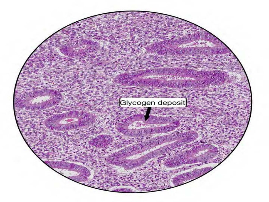
Among the given options, the histological image is likely to be of the endometrium. It shows the presence of glycogen masses in the basal cytoplasm of the epithelial cells lining the glands which are the features of the early secretory phase of the endometrium.
The uterus is made up of an external layer of smooth muscle called the myometrium, and an internal layer called the endometrium. The endometrium has three layers which are namely:
Stratum compactum,
Stratum spongiosum (which make up the stratum functionalis)
Stratum basalis.
The changes observed in the endometrium during the menstrual cycle are continuous and may be summarized as follows:
Increase in thickness of endometrium - The endometrium progressively increases in thickness.
In the postmenstrual phase, it is 0.5–1 mm thick; in the proliferative phase it is 2–3 mm thick, and in the secretory phase, its thickness reaches 5–7 mm.
Increase in dimensions of uterine glands - The uterine glands grow in length. At first, they are straight but gradually become convoluted. Because of these convolutions, the glands acquire a
'saw-toothed' appearance when seen in the longitudinal section. The glands also increase in
diameter.
• Change in the epithelial lining of glands - The epithelium lining the glands is at first cuboidal. During the proliferative stage, it becomes columnar. Glycogen accumulates in the basal portion of the epithelial cell, pushing the nucleus nearer the lumen. During the secretory phase, the apical part of the cell is shed off as part of the secretion and the cell again becomes cubical.
Given below is a histological section of the endometrium in the proliferative phase.
Q569.
Anatomy
Medium
4m
Image missing
Topic: Cardiovascular, Lymphatic and Respiratory SystemsSource: Internal
Explanation ready
Match the following marked structures to the correct areas of the lymph node.
Image not available for this question yet.
A) 1 - €, Å - A, ‚ - ƒ, „ - C, … - †
B) 1 - A, Å - ƒ, ‚ - †, „ - C, … - €
C) 1 - €, Å - ƒ, ‚ - A, „ - †, … -
D) 1 - A, Å - €, ‚ - †, „ - ƒ, … -
Correct Answer:C
Explanation:
The structures marked in the above histological section of the lymph node are as follows:
1 - Capsule
2 - Subcapsular sinus
3 - Germinal centre
4 - Lymphoid nodule
5 - Trabeculae
Each lymph node consists of a connective tissue framework and of numerous lymphocytes of all types which are arranged in a stroma of reticulin fibers and cells. They form 3 major regions within each lymph node:
Cortex - It contains subcapsular sinuses, lymphoid nodules, and densely packed lymphocytes.
It stains darkly. Each nodule has a lightly stained germinal center surrounded by a zone of densely packed lymphocytes.
Paracortex - It lacks nodules and has fewer lymphocytes. It is, therefore, lightly stained.
Medulla - with prominent draining sinusoids adjacent to the hilum.
Given below is a histological section of a lymph node showing the cortex, paracortex, and medulla.
Q570.
Anatomy
Medium
4m
Image missing
Topic: Cardiovascular, Lymphatic and Respiratory SystemsSource: Internal
Explanation ready
The arterioles present in the spleen divides into a number of straight vessels that are called
Image not available for this question yet.
A) Stave cells
B) €llipsoid
C) ‡enicilli
D) Ampulla
Correct Answer:C
Explanation:
The arterioles present in the spleen divide into a number of straight vessels that are called penicilli.
The splenic artery after entering the hilum of the spleen divides into about five branches that enter the organ independently. Each branch divides and subdivides as it travels through the trabecular network. Arterioles arising from this network leave the trabeculae to pass into the inter-trabecular spaces.
The spleen is histologically composed of:
White pulp - For some distance, each arteriole is surrounded by a dense sheath of lymphocytes.
These lymphocytes constitute the white pulp of the spleen.
Penicilli - The arteriole then divides into a number of straight vessels that are called
penicilli.
Ellipsoid - Each of the penicilli shows a localised thickening of its wall that is called an
ellipsoid.
The ellipsoid consists of concentric lamellae formed by aggregation of fibroblasts and macrophages.
Ampulla - Distal to the ellipsoid, the vessel dilates to form an ampulla, the walls of which become continuous with the reticular framework.
Red pulp - As a result, blood flows into spaces lined by reticular cells, coming into direct
contact with lymphocytes there. The part of splenic tissue, which is infiltrated with blood in this way is called the red pulp.
Sinusoids - Blood from spaces of the red pulp is collected by wide sinusoids that drain into veins in the trabeculae.
Stave cells: The sinusoids of the spleen are lined by a somewhat modified endothelium. The endothelial cells here are elongated and are shaped like bananas. They are referred to as stave cells.
Q571.
Anatomy
Medium
4m
Image missing
Topic: Cardiovascular, Lymphatic and Respiratory SystemsSource: Internal
Explanation ready
A mislabeled sample of tissue was sent over to the pathology lab for histopathological examination. On microscopy, a characteristic corpuscle as seen in the image was visualised. Which of the following structures is the sample most likely to be from?
Image not available for this question yet.
A) Spleen
B) Palatine tonsil
C) Thymus
D) Bone
Correct Answer:C
Explanation:
The sample is most likely to be of the thymus because of the presence of the corpuscle of Hassal as seen in the image above.
These are small rounded structures formed from eosinophilic type VI epithelial reticular cells present in the medulla of the thymus. Each corpuscle has a central core formed by
degenerated epithelial cells appearing as pink stained hyaline mass. Around this mass, there is a wall formed by concentrically arranged epithelial cells which stain bright pink in H and E staining.
Q572.
Anatomy
Medium
4m
Image missing
Topic: Cardiovascular, Lymphatic and Respiratory SystemsSource: Internal
Explanation ready
Given below is an image of a section of the small intestine. Identify the structure marked X 248
Image not available for this question yet.
A) Thymus
B) Spleen
C) MALT
D) Lymph node
Correct Answer:C
Explanation:
The structure marked X in the above given image of a section of ileum is the MALT (Mucosal associated lymphoid tissue), also known as gut-associated lymphoid tissue. These are seen as aggregated lymphatic follicle or Peyer's patch present in the submucosa of the ileum.
The mucosal associated lymphoid tissues are present in other regions as follows:
Tonsils - near the junction of the oral cavity with the pharynx, as collections of lymphoid
tissue.
The largest of these are the right and left palatine tonsils, present on either side of the oropharyngeal isthmus.
Solitary lymphatic follicles - small collections of lymphoid tissue, similar in structure to the follicles of lymph nodes, present anywhere along the length of the gut.
Aggregated lymphatic follicles (Peyer’s patches) - larger aggregations of lymphoid tissue, each consisting of 10 to 200 follicles present in the small intestine.
The image below of an ileal section shows Peyer's patches within the submucosa.
Q573.
Anatomy
Medium
4m
Image missing
Topic: Cardiovascular, Lymphatic and Respiratory SystemsSource: Internal
Explanation ready
Identify the structure shown in the histological image below. 249
Image not available for this question yet.
A) Trachea
B) Bronchus
C) Bronchiole
D) Alveolus
Correct Answer:A
Explanation:
The above histological image is that of the trachea as identified by the presence
of pseudostratified ciliated columnar epithelium lining, the serous and mucous glands and hyaline cartilage.
The histological section of the trachea shows the following structures:
Respiratory epithelium
Basement membrane
Submucosal glands (both serous and mucous parts)
Perichondrium
Tracheal cartilage
Trachealis muscle (smooth muscle).
The bronchi differ from the trachea in having plates rather than rings of cartilage, and in having a layer of smooth muscle between the lamina propria and submucosa. In smaller branches, the amount of cartilage decreases, whereas the amount of smooth muscles increases. Also, the number of glands and goblet cells decreases.
The image below is a histological section of the bronchus.
Q574.
Anatomy
Medium
4m
Image missing
Topic: Cardiovascular, Lymphatic and Respiratory SystemsSource: Internal
Explanation ready
The rounded secretory cells bearing microvilli present in the epithelial lining of the alveoli are
Image not available for this question yet.
A) Type I pneumocytes
B) Type II pneumocytes
C) Alveolar cells
D) Brush cells
Correct Answer:B
Explanation:
The secretory cells bearing microvilli present in the epithelial lining of the alveoli are the type II pneumocytes.
The cells forming the lining epithelium of alveoli (also called pneumocytes) are mainly of the following types:
Type I pneumocytes - large flattened cells which present a very thin diffusion barrier for gases.
They are connected to each other by tight junctions.
Type II pneumocytes - These are rounded secretory cells bearing microvilli on their free surfaces.
These cells secrete the surfactant which decreases the surface tension between the thin alveolar walls, and thus prevents alveoli from collapsing during respiration.
Type III alveolar cells (brush cells) - function is not clearly known.