Which of the following statements regarding the gastric glands is false?
Image not available for this question yet.
A) Pyloric antrum has many acid secreting cells
B) Oxyntic cells secrete hydrochloric acid
C) Cardiac region has many mucus secreting cells
D) Parietal cells secrete intrinsic factor
Correct Answer:A
Explanation:
The pylorus consists of mainly mucus-secreting glands.
The gastric glands can be divided into 3 groups which are namely:
Principal gastric glands - They are found in the body and fundus. About 3-7 principal glands open into the gastric pit. These glands contain at least 5 different types of cells:
Chief cells
Parietal cells
Mucous neck cells
Neuroendocrine cells
Stem cells.
Cardiac gastric glands - They are found in the small area near the cardiac orifice. Some are simple tubular glands, others are compound branched tubular. They contain mucus-secreting cells predominantly. Parietal and chief cells are sparsely present.
Pyloric gastric glands - Found in the pyloric antrum. About 2-3 convoluted tubes open into deep gastric pits. They mainly have mucus-secreting cells. They also have neuroendocrine cells, especially gastrin-secreting G cells. Parietal and chief cells are scarce.
Brunner's glands (or duodenal glands) are compound tubular submucosal glands present in the upper duodenum (above the sphincter of Oddi).
The alveoli of duodenal glands are lined mostly by mucous secreting columnar cells. Their ducts pass through the muscularis mucosae and empty into the intestinal crypts (of Lieberkuhn).
The duodenum can be easily differentiated from the jejunum or ileum by the presence of these submucosal glands. The jejunum and ileum do not have glands in their submucosa.
Given below is the histological section of normal duodenum showing the Brunner's glands at the submucosal layer.
An intern accidentally sent an incompletely labelled sample of the resected bowel wall from a patient with intestinal obstruction for histopathological examination. Given below is the microscopic image of the section of the sample. Which part of the gastrointestinal tract is being visualised here? 263
A) Esophagus
B) Stomach
C) Ileum
D) Duodenum
Correct Answer:C
Explanation:
The image is a section through the ileum as evident by the thin and slender villi and an aggregated lymphatic follicle (Peyer’s patch) in the submucosa.
These aggregations of lymphatic follicles are known as Mucosa Associated Lymphoid Tissue (MALT), and also as gut-associated lymphoid tissue (GALT). Peyer's patches in submucosa are found in large size and numerously in the terminal ileum.
In which of the following structures are centroacinar cells mainly present?
Image not available for this question yet.
A) Pancreas
B) Parotid gland
C) Prostate
D) Liver
Correct Answer:A
Explanation:
Centroacinar cells are mainly present in the pancreas.
Centroacinar cells are present in the alveoli of the exocrine pancreas. They are called so because they are visible near the centre of the acinus (or alveolus). These cells actually are actually part of the intercalated ducts that are invaginated into the secretory elements of the pancreas.
The space of Disse consists of all of the following except
Image not available for this question yet.
A) Microvilli
B) Blood plasma
C) Kupffer's cells
D) Fat cells
Correct Answer:C
Explanation:
The space of Disse consists of all of the following mentioned above except Kupffer cells. They are present in the lining of the walls of the sinusoids, not in the Space of Disse (perisinusoidal space).
Kupffer cells are also known as stellate macrophages or Kupffer-Borowicz cells. They are specialized macrophages present in the liver and are part of the mononuclear phagocyte system.
Space of Disse or the perisinusoidal space is present in the liver between the hepatocyte and a sinusoid. This space contains the blood plasma. The microvilli of hepatocytes extend into this space. This allows absorption of proteins and other plasma components from the sinusoids by the hepatocytes. This space may also have some fat cells.
The sinusoids are present in all the above-mentioned structures except kidneys. They are present in:
Liver
Spleen
Lymph nodes
Endocrine glands (adrenal medulla, parathyroid)
Bone
Carotid body
The given image is a graphical representation of sinusoids (sinus-like) which are small blood channels formed by subdivision of the lumen of large blood vessels (sinuses) by the invasion of developing parenchymal cell-cords.
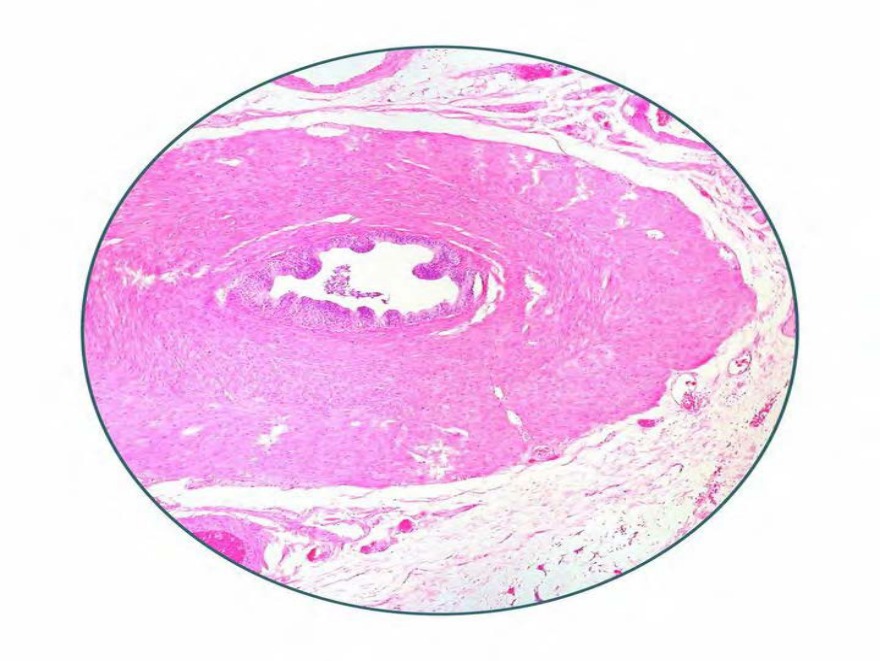
Identify the structure shown in the histological image below.
A) Female urethra
B) Male urethra
C) Ureter
D) Vas deferens
Correct Answer:C
Explanation:
The above image shows histological section of ureter.
The image shows a thick, fibroelastic, lamina propria lying underneath the stratified, transitional epithelium. There are no mucosal or submucosal glands and no submucosa. There is a layer of smooth muscle outside the mucosa. The outer adventitial layer has fibroelastic connective tissue, with blood vessels, lymphatics and nerves.
Given below is a cross-section of the ureter showing the transitional epithelium and the layer of smooth muscle.
Q583.
Anatomy
Medium
4m
Image missing
Topic: Skin Special Senses, Eye and EarSource: Internal
Explanation ready
Match the following marked structures to the correct layers of the epidermis.
Image not available for this question yet.
A) A-3, B-4, C-2, D-1
B) A-3, B-2, C-4, D-1
C) A-2, B-4, C-3, D-1
D) A-2, B-1, C-3, D-4
Correct Answer:D
Explanation:
The image shows the histological section of the epidermis. The structures marked are respectively:
Stratum corneum
Stratum granulosum
Stratum spinosum
Stratum basale
The above 4 layers are present in the thin hairy skin.
The thick glabrous skin over the palms and the soles alone contains a clear homogenous 5th layer between the stratum corneum and stratum granulosum, called the stratum lucidum.
Q584.
Anatomy
Medium
4m
Image missing
Topic: Skin Special Senses, Eye and EarSource: Internal
Explanation ready
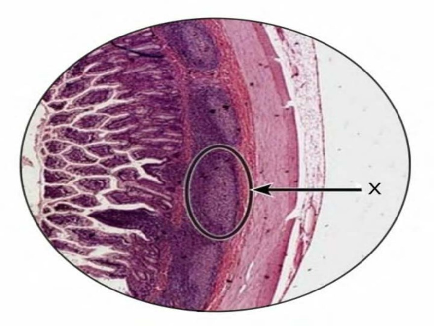
Given below is a histological section showing the layers of the skin. The region marked X points to the location of
Image not available for this question yet.
A) Melanocytes
B) Duct of sweat glands
C) Desmosomes
D) Hemidesmosomes
Correct Answer:C
Explanation:
Among the given options, the region marked X points towards the location of intercellular adhesions formed by desmosomes.
Desmosomes (maculae adherens) are specialized adhesive protein complexes that localize to intercellular junctions. They are responsible for maintaining the mechanical integrity of tissues and resisting shearing forces. They are made of intercellular filaments, dense cytoplasmic plaques within the cells on either side and intermediate filaments.
Hemidesmosomes are specialized structures for the cell to extracellular material adhesions.
The schematic representation shows the desmosomal proteins and relative distance from the plasma membrane. The desmosomal cadherins, the desmogleins and desmocollins, extend into the intercellular space and outer dense plaque to establish contact and adhere to neighbouring cells. The cadherin cytoplasmic tails associate linker proteins, plakoglobin, plakophilins, and desmoplakin. The desmoplakin binds to keratin intermediate filaments within the inner dense plaque, serving to tether the intermediate filaments to the plasma membrane.
Q585.
Anatomy
Medium
4m
Image missing
Topic: Skin Special Senses, Eye and EarSource: Internal
Explanation ready
Given below is an image of a histological section of the skin. Identify the structure marked X. 289
Image not available for this question yet.
A) Merkel cell
B) Meissner corpuscles
C) Pacinian corpuscles
D) Ruffini’s corpuscles
Correct Answer:B
Explanation:
The structure marked X in the histological image of the skin is the Meissner's corpuscle which is located in the papillary dermis.
Each corpuscle consists of a connective tissue capsule and a central core composed of a stack of flat modified Schwann cells (Coiled spring-like morphology). They are present in the dermal papillae of all parts of hands and foot, anterior aspect of the forearm, lips and mucous membrane of the apical part of the tongue.
They are rapidly acting mechanoreceptors and are specialised for light touch and low-frequency stimuli. They are sensitive to shape and textural changes and provide the neural basis for reading Braille text.
The histological image below shows Meissner's corpuscles in the dermal papillae.
Q586.
Anatomy
Medium
4m
Image missing
Topic: Skin Special Senses, Eye and EarSource: Internal
Explanation ready
Panniculus adiposus is present in:
Image not available for this question yet.
A) Axilla
B) Scrotum
C) Eyelid
D) Penis
Correct Answer:A
Explanation:
Panniculus adiposus is present in the axilla.
Panniculus adiposus refers to a layer/sheet of fat present subcutaneously and is found over most of the body except the scrotum, penis, and eyelids. It is responsible for giving a smooth contour to the skin.
Q587.
Anatomy
Medium
4m
Image missing
Topic: Skin Special Senses, Eye and EarSource: Internal
Explanation ready
Arrange from outer to inner the layers of the retina given below: A-Retinal pigment epithelium B-Inner plexiform layer C-Nerve fiber layer D-Inner nuclear layer E-external limiting membrane.
Image not available for this question yet.
A) E,D,C,B,A
B) A,B,D,C,E
C) A,E,D,B,C
D) E,A,D,B,C
Correct Answer:C
Explanation:
From the given options, the correct sequence from outer to inner is:
retinal pigment epithelium (A),
external limiting membrane (E),
inner nuclear layer (D),
inner plexiform layer (B),
nerve fiber layer (C)
Q588.
Anatomy
Medium
4m
Image missing
Topic: Skin Special Senses, Eye and EarSource: Internal
Explanation ready
The gelatinous component of the crista ampullaris that extends from the crista to the roof of the ampullae is known as
Image not available for this question yet.
A) Calix
B) Spiral ganglion
C) Cupula
D) Spiral ligament
Correct Answer:C
Explanation:
The gelatinous component of the crista ampullaris that extends from the crista to the roof of the ampullae is known as the cupula.
The ampullary cupula, or cupula, provides a sense of spatial orientation. It is located in each of the ampullae of the three semicircular canals. In the crista ampullaris, hair cells that have several stereocilia associated with each kinocilium, are embedded into the gelatinous matrix of the cupula in a specific pattern that is functionally significant. The lag of endolymph on the rotational movement of the head deflects the cupula and hence stimulates the hair cells.
The pattern of the arrangement of cranial nerve nuclei from the ventral to dorsal aspect is: Somatic Efferent gt; Visceral Efferent gt; Visceral Afferent gt; Somatic Afferent
The basal and alar cranial nerve nuclei of the brainstem are organized into 7 columns. The sequence of the columns from ventral to dorsal is shown in the image below - 3 motor columns in the ventral basal plate (green) and 4 sensory columns in the dorsal alar plate (blue). The visceral nuclei are closer to the sulcus limitans than the somatic nuclei.
A patient with trigeminal neuralgia presents with a flare-up of the condition. The nucleus receiving the sensory afferent extends from the chief sensory nucleus in the pons up to the .
Image not available for this question yet.
A) Lower end of medulla
B) 2nd cervical spinal segment
C) 5th cervical spinal segment
D) Upper end of medulla
Correct Answer:B
Explanation:
Excruciating pain is the principal symptom of trigeminal neuralgia. The afferent fibers carrying pain and temperature from the face relay in the spinal nucleus of the trigeminal nerve. It extends from the chief sensory nucleus in the upper part of the pons through the medulla and into the upper 2 segments of the spinal cord.
The trigeminal nerve consists of 4 nuclei: 3 sensory (mesencephalic, spinal, and principal sensory nucleus) and 1 motor.
Nucleus Mesencephalic nucleus
Principal sensory nucleu s
Spinal nucleus
Motor nucleus
Location
Composed of a column ofuni polar nervecells situated in th e midbrain and extends inferi orly into the pons as far as th e main sensory nucleus
Posterior part of the pons lat eral to the motor nucleus
Continuous superiorly with t he main sensory nucleus and extends inferiorly the whole of the medulla oblongatatill t he 2ndcervical segment
The pons medial to the main sensory nucleus
Function
Receives the sensation ofpro prioceptionfrom the muscles of mastication and from the f acial and extraocular muscles
Receives the sensations oftou ch and pressurefrom the face
Receives the sensation ofpain and temperaturefrom the fac e and some fibers oftouch
Supplies the muscles of masti cation, tensor tympani, tenso r veli palitini, mylohyoid, and anterior belly of digastric m uscle
Q591.
Anatomy
Medium
4m
Image missing
Topic: Meninges and dural venous sinusesSource: Internal
Explanation ready
What is the nerve supply of the dura covering the marked region? 328
Image not available for this question yet.
A) C2-C3 via cranial nerves X and XII
B) Anterior and posterior ethmoidal nerves
C) Nervi meningeus medius and spinosus
D) Cranial nerves VII and IX only
Correct Answer:A
Explanation:
The dura mater of the posterior cranial fossa is mainly innervated by C2 and C3 nerves via cranial nerves X and XII.
These fibers may be distributed by the vagus and hypoglossal nerves.
Q592.
Anatomy
Medium
4m
Image missing
Topic: Ventricular System and Subarachnoid SpaceSource: Internal
Explanation ready
The CT scan of a child with congenital toxoplasmosis is shown below. Which of the following cells form the lining of the involved structure?
Image not available for this question yet.
A) Ependymocytes
B) Astrocytes
C) Oligodendrocytes
D) Podocytes
Correct Answer:A
Explanation:
The CT shows hydrocephalus, enlargement of the ventricles due to build-up of CSF. The ventricles of the brain are lined by ependymocytes.
Ependymocytes are a type of glial cell that line the ventricles of the brain and the central canal of the spinal cord. They are simple columnar cells. Some are ciliated in order to facilitate the circulation of CSF.
Their functions include secretion of CSF and transport of neurochemicals to and fro from the CSF. They also have a function in neuroregeneration.
Q593.
Anatomy
Medium
4m
Image missing
Topic: Ventricular System and Subarachnoid SpaceSource: Internal
Explanation ready
What structure forms the roof of the anterior horn of the marked ventricle? 341
Image not available for this question yet.
A) Genu of corpus callosum
B) Rostrum of corpus callosum
C) Body of corpus callosum
D) Septum pellucidum
Correct Answer:C
Explanation:
The structure marked is the lateral ventricle. The roof of the anterior horn of the lateral ventricle is formed by the anterior part of the body of the corpus callosum.
The lateral ventricle is divided into the body and 3 horns. The locations of the parts are:
Body - parietal lobe
Anterior horn - frontal lobe
Posterior horn - occipital lobe
Inferior horn - temporal lobe.
The anterior horn extends from the genu of the corpus callosum up to the interventricular foramen. The relations of the anterior horn are:
Anteriorly - Genu and rostrum of the corpus callosum
Roof - Anterior part of the body of the corpus callosum
Floor and lateral wall - Caudate nucleus
Medial wall - Septum pellucidum and anterior column of fornix
The body or central part extends from the interventricular foramen to the splenium of the corpus callosum. The relations of the body are:
Roof - Body of the corpus callosum
Medial wall - Septum pellucidum
Floor (from lateral to medial) - Caudate nucleus, stria terminalis, thalamostriate vein, thalamus, choroid plexus, and body of the fornix
The posterior horn extends from the splenium to the occipital pole. The relations are:
Roof and lateral wall - Tapetum of corpus callosum
Medial wall - Forceps major and calcar avis (calcarine sulcus)
The inferior horn is the largest compartment of the lateral ventricle and extends up to 2.5 cm from the temporal pole. The relations are:
Roof - Tapetum of corpus callosum and tail of caudate nucleus
Floor (lateral to medial) - Collateral eminence produced by collateral fissure, hippocampus.
Note: There is no lateral wall for the lateral ventricle.
Q594.
Anatomy
Medium
4m
Image missing
Topic: Ventricular System and Subarachnoid SpaceSource: Internal
Explanation ready
The two elevations on the medial wall of the posterior horn of the lateral ventricle are due to .
Image not available for this question yet.
A) Tapetum of the corpus callosum and calcar avis
B) Forceps minor and collateral eminence
C) Forceps major and calcar avis
D) Forceps major and collateral eminence
Correct Answer:C
Explanation:
The two elevations on the medial wall of the posterior horn of the lateral ventricle are due to the forceps major and calcar avis.
The posterior horn extends from the splenium to the occipital pole. The relations are:
Roof and lateral wall - Tapetum of corpus callosum
Medial wall - Forceps major and calcar avis (calcarine sulcus)
The forceps major is the fibers of the splenium of the corpus callosum. The calcar avis is formed by the white matter of the calcarine sulcus.
Forceps minor is formed by the anterior part of the corpus callosum.
The collateral eminence is formed by the collateral sulcus and is related to the floor of the inferior horn of the lateral ventricle.
Q595.
Anatomy
Medium
4m
Image missing
Topic: Ventricular System and Subarachnoid SpaceSource: Internal
Explanation ready
The T2 weighted MRI of a child is given below. Which of the following is true about the ventricle primarily involved in this condition?
Image not available for this question yet.
A) Foramen of Magendie is a central aperture that pierces the superior medullary velum
B) Inferior medullary velum is a sheet of white matter
C) Foramina of Luschka are the lateral openings
D) The choroid plexus is formed from folds of arachnoid called tela choroidea
Correct Answer:C
Explanation:
The MRI shows a Dandy-Walker malformation. It is a rare congenital malformation involving the cerebellum and the fourth ventricle. The foramina of Luschka are the lateral openings in the fourth ventricle. These openings allow the flow of CSF from the ventricle into the subarachnoid space.
The fourth ventricle is a tent-shaped ventricle lying between the brainstem and the cerebellum. It is widest at the level of the pontomedullary junction.
The roof of the fourth ventricle is formed by:
Superior medullary velum - White matter sheet stretching between the superior cerebellar peduncles.
Inferior medullary velum - Made of ventricular ependyma and pia mater of tela choroidea, and has no nervous tissue. It is pierced by the foramen of Magendie in the midline, opening into the cisterna magna.
The T-shaped choroid plexus of the fourth ventricle is formed from the tela choroidea, made of two layers of pia mater.
Q596.
Anatomy
Medium
4m
Image missing
Topic: Ventricular System and Subarachnoid SpaceSource: Internal
Explanation ready
The structure marked with arrow in the image given below is concerned with .
Image not available for this question yet.
A) Secretion of the CSF into the subarachnoid space
B) Transport of neurochemicals from subjacent neurons and glia into CSF
C) Absorption of the CSF by the arachnoid villi
D) CSF return through the tela choroidea
Correct Answer:C
Explanation:
The structure marked in the above image is a dural venous sinus, and the CSF is absorbed into this sinus through arachnoid villi.
Tela choroidea gives rise to the choroid plexus, from which CSF is secreted into the ventricles.
Tancytes/specialized glial cells in circumventricular organs help with the transport of neurochemicals to CSF.
Q597.
Anatomy
Medium
4m
Image missing
Topic: Ventricular System and Subarachnoid SpaceSource: Internal
Explanation ready
While assessing a 33-year-old lady who had been in a road traffic accident, you note watery discharge from her nose mixed with the blood. The following sign is seen on filter paper. Which structures produce the majority of this fluid?
Image not available for this question yet.
A) Ependymal cells lining the ventricles
B) Choroid plexus
C) Brain substance through perivascular space
D) Microglia
Correct Answer:B
Explanation:
The case suggests CSF rhinorrhea and the image shows the double-ring sign. CSF mainly forms in the choroid plexus. It is absorbed back by the arachnoid villi.
Formation of CSF:
Major contributor - Choroid plexuses
Minor contributors - Ependymal cells lining the ventricles, brain substance through the perivascular spaces
CSF is produced at a rate of 55– ml/day. The normal adult volume of CSF is 15–ml, and it is cycled about 3.˜ times a day. The normal CSF pressure is ˜–-1™– mm H2O.
Q598.
Anatomy
Medium
4m
Image missing
Topic: Ventricular System and Subarachnoid SpaceSource: Internal
Explanation ready
Which of the following is not a circumventricular organ? 347
Image not available for this question yet.
A) Organum Vasculosum
B) Median Eminence
C) Adenohypophysis
D) Pineal Gland
Correct Answer:C
Explanation:
Circumventricular organs are those structures in the brain that are devoid of the blood-brain barrier. Adenohypophysis is not devoid of a blood-brain barrier (BBB).
Neurohypophysis is devoid of the blood-brain barrier.
Q599.
Anatomy
Medium
4m
Image missing
Topic: White Matter of the BrainSource: Internal
Explanation ready
An infant is referred to neurology due to an abnormal MRI report as shown below. What is the most anterior part of the missing brain structure?
Image not available for this question yet.
A) Rostrum
B) Genu
C) Splenium
D) Trunk
Correct Answer:B
Explanation:
The given clinical scenario is suggestive of total agenesis of the corpus callosum. The most anterior part of the missing brain structure i.e. the corpus callosum is the genu.
The image given below shows an MRI scan of the sagittal section of a normal brain followed by an image showing corpus callosum agenesis.
The correct order of arrangement of stem cells, based on their potency to differentiate into different cells (highest to lowest) is totipotent gt; pluripotent gt; multipotent gt; oligopotent gt; unipotent.