The structure marked is the membranous part of the interventricular septum. It is derived from endocardial cushions.
Endocardial cushions contribute to the formation of:
Atrial septa
Ventricular septa (membranous portion)
Atrioventricular canals and valves
Aortic and pulmonary channels
Q1602.
Anatomy
Medium
4m
Image missing
Topic: HeartSource: Internal
Explanation ready
All of the following supply the marked structure except :
A) Right coronary artery
B) Anterior interventricular artery
C) Marginal artery
D) Diagonal artery
Correct Answer:D
Explanation:
The structure marked is the right ventricle. It is not supplied by the diagonal artery.
The diagonal artery is a branch of the anterior interventricular artery supplying the left ventricle. This is present in about 30-50 of the population.
The right ventricle is mostly supplied by the right coronary artery and its branches such as the marginal artery.
The anterior interventricular artery gives off 1-2 anterior right ventricular branches.
What is the nerve supply of the structure marked in the image below?
Image not available for this question yet.
A) Posterior interosseus nerve
B) Median nerve
C) Anterior interosseus nerve
D) ’lnar nerve
Correct Answer:B
Explanation:
The marked structure is second lumbrical which is supplied by the median nerve.
The lateral two lumbricals are supplied by the median nerve and the medial two are supplied by the ulnar nerve. Function of these muscles is flexion at the metacarpophalangeal joints and extension at both the interphalangeal joints.
Q1604.
Anatomy
Medium
1mPYQ
Image missing
Topic: AIIMS 2020 (May and Nov INI-CET)Source: AIIMSYear: 2020
Explanation ready
Given is a model of embryological development of the heart. Which structure develops from the area marked as "A" ?
Image not available for this question yet.
A) Atria
B) Left Ventricle
C) Infundibulum
D) Interventricular septum
Correct Answer:C
Explanation:
The structure marked is conus cordis (middle part of bulbus cordis). The infundibulum of the heart develops from this structure.
The model shows the embryologic stage of the heart at which both the heart tubes have fused and are undergoing dextro-looping or bending. This happens around day 23 of intrauterine life.
Q1605.
Anatomy
Medium
1mPYQ
Image missing
Topic: AIIMS 2020 (May and Nov INI-CET)Source: AIIMSYear: 2020
Explanation ready
A muscle is attached to the area marked in the given image. What is the action of this muscle on the hip joint?
Image not available for this question yet.
A) Flexion and medial rotation
B) Adduction and lateral rotation
C) Abduction and medial rotation
D) Extension and lateral rotation
Correct Answer:D
Explanation:
The area marked in the given image is the gluteal tuberosity. The muscle that is inserted here is the gluteus maximus which causes extension and lateral rotation of the hip joint.
Gluteus maximus inserts onto the iliotibial tract (3/4th part) and deep fibers (1/4th) attach on the gluteal tuberosity. It is supplied by the inferior gluteal nerve.
Q1606.
Anatomy
Hard
1mPYQ
Image missing
Topic: AIIMS 2020 (May and Nov INI-CET)Source: AIIMSYear: 2020
Explanation ready
Neurotransmitter secreted by the area marked in the given image is?
Image not available for this question yet.
A) Acetylcholine
B) Dopamine
C) Glutamine
D) Noradrenaline
Correct Answer:B
Explanation:
The area marked in the given image is substantia nigra (meaning- dark substance). The neurotransmitter secreted by this area is dopamine.
Substantia nigra is a part of the nigrostriatal pathway. Degeneration of dopaminergic neurons is implicated in Parkinson's disease.
Q1607.
Anatomy
Medium
1mPYQ
Image missing
Topic: AIIMS 2020 (May and Nov INI-CET)Source: AIIMSYear: 2020
Explanation ready
Which of the following arteries passes in close proximity to the area marked by the arrow in the given image?
Image not available for this question yet.
A) Internal carotid artery
B) Middle meningeal artery
C) Anterior cerebral artery
D) Deep temporal artery
Correct Answer:B
Explanation:
The area marked by an arrow in the given image is pterion and the artery that passes in close proximity to this area is the middle meningeal artery.
The pterion is an H-shaped sutural junction between four bones of the skull: Frontal bone, Squamous part of the temporal bone, Greater wing of the sphenoid bone, and Parietal bone. It overlies the anterior branch of the middle meningeal artery. Rupture of this artery can lead to an extradural hematoma.
Q1608.
Anatomy
Medium
1mPYQ
Image missing
Topic: AIIMS 2020 (May and Nov INI-CET)Source: AIIMSYear: 2020
Explanation ready
Where are the Ito cells in the liver located?
Image not available for this question yet.
A) Space of disse
B) Space of moll
C) Sinusoids
D) Bile canaliculi
Correct Answer:A
Explanation:
Ito cells/Hepatic stellate cells are located in the space of Disse. They are mesenchymal in origin.
The space of Disse or the perisinusoidal space is a location in the liver between hepatocytes and the liver sinusoid. Ito cells store fat-soluble vitamin A in their lipid droplets.
Q1609.
Anatomy
Hard
1mPYQ
Image missing
Topic: AIIMS 2020 (May and Nov INI-CET)Source: AIIMSYear: 2020
Explanation ready
Which of the following features differentiates the exocrine pancreas from the parotid gland?
Image not available for this question yet.
A) Basal lamina lining the serous acini
B) Absence of striated duct
C) Presence of serous acini with acidophilic tip
D) Presence of apical acinar villi
Correct Answer:B
Explanation:
Absence of striated duct in the exocrine pancreas differentiates it from the parotid gland.
The exocrine pancreas is comprised entirely of serous acini. Its general structure is similar to that of the parotid gland, but the parotid gland has striated and intercalated intralobular ducts, whereas the pancreas has only the intercalated type.
Q1610.
Anatomy
Hard
1mPYQ
Image missing
Topic: AIIMS 2020 (May and Nov INI-CET)Source: AIIMSYear: 2020
Explanation ready
Medial lemniscus is a continuation of which of the following structures?
Image not available for this question yet.
A) Spinothalamic tract
B) Spinocerebellar tract
C) Spinotectal tract
D) Fasciculus gracilis
Correct Answer:D
Explanation:
Medial lemniscus is a continuation of fasciculus gracilis.
Fibers of fasciculus gracilis and cuneatus synapse on the second-order neurons of the ipsilateral nuclei gracilis and cuneatus in the medulla oblongata. Axons of these second-order neurons then cross over as the internal arcuate fibers to form the medial lemniscus in the contralateral medulla.
Q1611.
Anatomy
Hard
1mPYQ
Image missing
Topic: AIIMS 2020 (May and Nov INI-CET)Source: AIIMSYear: 2020
Explanation ready
The type of fibers present in the fornix are:
Image not available for this question yet.
A) Association and commisural fibers
B) Association and projection fibers
C) Commissural and projection fibers
D) Association fibers
Correct Answer:A
Explanation:
The fibers present in the fornix are association fibers except for the hippocampal commissure of the fornix which has commissural fibers.
Association fibers connect various cortical regions within the same hemisphere whereas commissural fibers connect corresponding regions of two hemispheres. Fornix arises from the hippocampus and ends in the mamillary body of the hypothalamus.
Q1612.
Anatomy
Medium
1mPYQ
Image missing
Topic: AIIMS 2020 (May and Nov INI-CET)Source: AIIMSYear: 2020
Explanation ready
Decussation of the superior cerebellar peduncle occurs at which level?
Image not available for this question yet.
A) Pons
B) Midbrain
C) Diencephalon
D) Medulla
Correct Answer:B
Explanation:
The decussation of the superior cerebellar peduncle occurs in the midbrain, at the level of the inferior colliculus.
The midbrain has four decussations. At the level of the inferior colliculus: Decussation of superior cerebellar peduncles and decussation of trochlear nerve.
Q1613.
Anatomy
Easy
1mPYQ
Image missing
Topic: AIIMS 2020 (May and Nov INI-CET)Source: AIIMSYear: 2020
Explanation ready
The pretracheal layer of deep cervical fascia encloses all of the following except:
Image not available for this question yet.
A) Oesophagus
B) Sternocleidomastoid
C) Sternohyoid
D) Thyroid gland
Correct Answer:B
Explanation:
All the above structures are enclosed by the pretracheal layer of deep cervical fascia except sternocleidomastoid.
Sternocleidomastoid is enclosed by the investing layer of deep cervical fascia. The pretracheal fascia covers the strap muscles (sternohyoid, omohyoid, etc.), splits to enclose the thyroid gland, and surrounds the trachea and the esophagus.
Q1614.
Anatomy
Easy
1mPYQ
Image missing
Topic: AIIMS 2020 (May and Nov INI-CET)Source: AIIMSYear: 2020
Explanation ready
All of the following are the contents of the carotid sheath except?
Image not available for this question yet.
A) Sympathetic chain
B) Carotid artery
C) Internal jugular vein
D) 10th cranial nerve
Correct Answer:A
Explanation:
The sympathetic chain is not the content of the carotid sheath.
It lies posterior to the carotid sheath and anterior to the transverse processes of the cervical vertebrae in the neck. Contents of carotid sheath: Common and internal carotid arteries, Internal jugular vein, Vagus nerve (10th CN), and Ansa cervicalis.
A patient presented with paradoxical respiration. Which of the following nerves is most likely to be affected?
Image not available for this question yet.
A)
B) B
C) C
D) D
Correct Answer:D
Explanation:
Paradoxical respiration occurs due to the damage of the phrenic nerve, which is marked as D, in the given image.
The structures marked in the image are:
A. Right vagus nerve
B. Left vagus nerve
C. Recurrent laryngeal nerve
D. Right phrenic nerve
Paradoxical movements in the chest are as follows:
Paradoxical respiration - the abnormal movement of the chest wall or abdominal wall, where they move in during inspiration instead of moving out. It is seen in flail chest.
Paradoxical movement of the diaphragm - normally, the diaphragm moves down during inspiration. However, in cases of phrenic nerve palsy, the affected side of the diaphragm moves upward (paradoxical respiration) due to diaphragmatic paralysis. The gold standard investigation to detect this is the electrical or magnetic stimulation of the phrenic nerve with a recording of
the compound muscle action potential and/or the transdiaphragmatic pressure. This can be observed on ultrasound or fluoroscopy.
Which of the following muscles is attached to the area marked in the image below?
Image not available for this question yet.
A) Abductor pollicis longus
B) Palmar interossei
C) Opponens pollicis
D) Flexor pollicis brevis
Correct Answer:C
Explanation:
The area marked in the image is the palmar aspect of the first metacarpal. The opponens pollicis muscle is inserted at this site.
Opponens pollicis:
Origin: Tubercle of the trapezium and flexor retinaculum
Insertion: Whole length of the lateral border and adjoining lateral half of the palmar aspect of the first metacarpal.
Action: Flexes the metacarpal bone of the thumb.
The image below shows various intrinsic muscles of the hand.
Q1617.
Anatomy
Medium
1mPYQ
Image missing
Topic: AIIMS 2020 (May and Nov INI-CET)Source: AIIMSYear: 2020
Explanation ready
Which of the following marked structures does not form part of the laryngeal skeleton?
Image not available for this question yet.
A) A
B) B
C) C
D) D
Correct Answer:A
Explanation:
The structure marked as 'A', is the hyoid bone, which is not a part of the laryngeal skeleton.
The laryngeal skeleton is formed by a series of cartilages that are connected by ligaments and fibrous membranes. The structures marked in the image are: A - Hyoid bone, B - Thyroid cartilage, C - Epiglottis, D - Cricoid cartilage.
Q1618.
Anatomy
Medium
4m
Image missing
Topic: Osteology, Scalp and FaceSource: Internal
Explanation ready
Identify the foramen marked in the image below.
Image not available for this question yet.
A) ‡oramen rotundum
B) ‡oramen ovale
C) ˆidian canal
D) ‡oramen spinosum
Correct Answer:A
Explanation:
The image shows the sphenoid bone. The foramen marked is the foramen rotundum. Maxillary branch of trigeminal nerve passes through it.
Reticular fibres are not present in which of the following structures?
Image not available for this question yet.
A) Thymus
B) Lymph node
C) Bone
D) Spleen
Correct Answer:A
Explanation:
Reticular fibres are not present in the thymus.
Reticular fibres are fine, anastomosing fibres composed of mainly type III collagen. They differ from type I collagen fibres in that, they are finer and are arranged in the form of a network (reticulum). The fibres appear black in colour on staining with silver impregnation. Due to this affinity to silver salts, they are also known as argentophil fibres.
Reticular fibres are mainly found in:
All lymphoid organs like spleen, lymph node, and bone (except thymus).
Which of the following muscles is not involved in the overhead abduction of the arm?
Image not available for this question yet.
A) Serratus anterior
B) Deltoid
C) Trapezius
D) Pectoralis major
Correct Answer:D
Explanation:
Pectoralis major is mainly involved in adduction and medial rotation of the arm and is not involved in the abduction of the arm.
Glenohumeral abduction is about 120° or more. The glenohumeral arthrodesis restricts the abduction at about 45°. Further abduction occurs at the sterno and acromioclavicular articulations, and contralateral vertebral flexion.
Muscles involved in the abduction of the shoulder:
The first 15° of abduction is initiated by the supraspinatus muscle.
15° to 90° of abduction is carried out mainly by the middle fibers of the deltoid muscle.
Abduction of the arm, beyond 90°, is carried out by the movement of the shoulder girdle caused by the action of trapezius and serratus anterior.
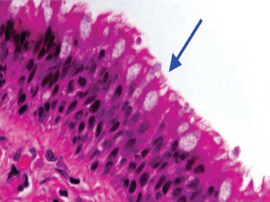
The structures marked in the image below are found in the epithelial lining of all of the following sites, except:
A) Oviduct
B) Epididymis
C) Bronchi
D) Ependyma
Correct Answer:B
Explanation:
The image shows an arrow mark pointing towards the cilia. All the structures given in the options have a ciliated lining epithelium except epididymis, which is lined by pseudostratified columnar epithelium with stereocilia.
The given image is a histological section of the epididymis showing the pseudostratified columnar epithelium with stereocilia.
Cilia are minute hair-like projections, while stereocilia are very long, thick microvilli measuring about 5–10 μm in length.
Ciliated epithelium:
The epithelium bears cilia, which moves in a coordinated fashion, and help in the movement of the mucus, or propel the movement of any other structures such as ova or spermatozoa.
The ciliated columnar epithelium is found in most of the respiratory tract, the uterus, and the uterine tube (oviduct), efferent ductules of the testis, parts of the middle ear and auditory tube;
the ependyma lining the central canal of the spinal cord and the ventricles of the brain.
The pseudostratified ciliated columnar epithelium is seen in the trachea and in large bronchi.
Q1622.
Anatomy
Medium
4m
Image missing
Topic: Osteology, Scalp and FaceSource: Internal
Explanation ready
Which of the following bony structures is present behind the marked area?
Image not available for this question yet.
A) Post clivus
B) Lesser wing of sphenoid
C) Sella turcica
D) Carotid canal
Correct Answer:B
Explanation:
The structure marked in the image is pterion, and the bony structure that is present behind it is the lesser wing of the sphenoid.
The pterion is an H-shaped sutural junction between four bones of the skull:
Frontal bone
Squamous part of the temporal bone
Greater wing of the sphenoid bone
Parietal bone
The pterion overlies the anterior branch of the middle meningeal artery and lateral fissure of the cerebral hemisphere. The bone in this area is thin and any injury to this area may lead to rupture of the middle meningeal artery leading to extradural hematoma.
The pterion also corresponds to the site of the anterolateral (sphenoidal) fontanelle of the neonatal skull, which closes in the third month after birth.
All of the following nerves are involved in the movement of the eyeball except:
Image not available for this question yet.
A)
B) B
C) C
D) D
Correct Answer:A
Explanation:
The nerve marked as 'A' is the optic nerve (II CN), and is not involved in the movement of the eyeball.
The nerves marked in the image are:
Optic nerve (II CN)
Oculomotor nerve (III CN)
Abducens nerve (VI CN)
Trochlear nerve (IV CN)
Extraocular muscles involved in the movement of the eyeball:
Superior rectus, inferior rectus, medial rectus, inferior oblique, and levator palpebrae
superioris muscles - supplied by the oculomotor (III) nerve.
Lateral rectus - supplied by abducens (VI) nerve.
Superior oblique - supplied by trochlear (IV) nerve.
Q1624.
Anatomy
Medium
4m
Image missing
Topic: Upper Limb Bones and JointsSource: Internal
Explanation ready
The middle radioulnar joint is an example of a:
Image not available for this question yet.
A) Plane synovial
B) Primary cartilaginous
C) Secondary cartilaginous
D) Fibrous joint
Correct Answer:D
Explanation:
The middle radioulnar joint is an example of a fibrous joint (syndesmosis).
The radius and ulna are united with each other by three joints:
Proximal (superior) radioulnar joint
Middle radioulnar joint
Distal (inferior) radioulnar joint
Proximal and distal radioulnar joints are uniaxial pivot joints (synovial) and are involved in pronation and supination movements of the forearm.
The middle radioulnar joint is a fibrous joint (syndesmosis). The radius and ulna, in the middle, are connected by two syndesmoses namely:
The oblique cord - extends from the lateral side of the ulnar tuberosity to the radius, distal to
the radial tuberosity.
Interosseous membrane - a broad, thin collagenous structure, which runs perpendicular to the oblique cord. It extends distomedially between the interosseous borders of the radius and ulna.
Based on the structure, joints are classified into three types:
Fibrous joints
Sutures
Syndesmosis
Gomphosis
Cartilaginous joints
Primary cartilaginous joints or synchondrosis
Secondary cartilaginous joints or symphysis
Synovial joints
Ball-and-socket or spheroidal joints
Sellar or saddle joints
Condylar or bicondylar joints
Ellipsoid joints
Hinge joints
Pivot or trochoid joints
Plane joints
Q1625.
Anatomy
Medium
4m
Image missing
Topic: Muscles of the Lower LimbSource: Internal
Explanation ready
What is the action of the muscle at the hip, which is attached to the marked area in the image below.
Image not available for this question yet.
A) Abduction and external rotation
B) Extension and lateral rotation
C) Extension, adduction, and lateral rotation
D) Flexion and abduction
Correct Answer:B
Explanation:
The image shows the posterior surface of the femur, and the structure marked is gluteal tuberosity, to which the gluteus maximus muscle is attached. It causes extension and lateral rotation at the hip joint.
The femur can be divided into:
The proximal femur - Consists of the head, neck, greater and lesser trochanters. The intertrochanteric line is present on the anterior surface at the junction of the neck and shaft of
the femur while the intertrochanteric crest is present on the posterior surface.
The shaft of the femur - Consists of linea aspera (bony crest present on the posterior surface), gluteal tuberosity, and pectineal line
The distal femur - Consists of the medial condyle, lateral condyle, intercondylar fossa, and patellar surface
The image below shows attachments of various muscles over the posterior surface of the right femur.
Gluteus maximus muscle:
Origin:
Posterior aspect of the dorsal ilium, posterior to the posterior gluteal line
Posterior superior iliac spine
Posterior aspect of sacrum and coccyx
Sacrotuberous ligament.
Insertion - It is inserted mainly in fascia lata at the iliotibial band and also into the gluteal tuberosity on the posterior femoral surface.
Action:
Major extensor of the hip joint
Lateral rotation of the hip joint
Active in powerful abduction of the thigh
Arterial Supply - Inferior and superior gluteal arteries and the first perforating branch of the profunda femoris artery.
Nerve Supply- Inferior gluteal nerve (L5, S1 and S2).